Scientists discover the body’s hidden “off switch” for inflammation

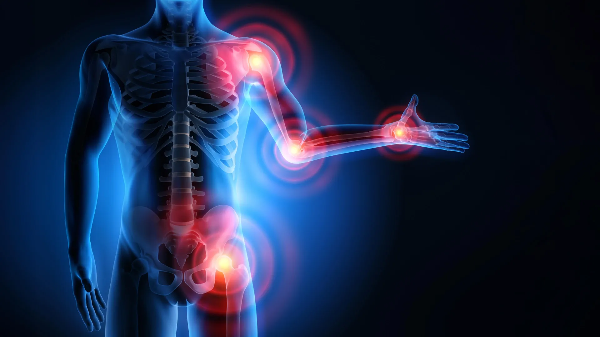
Researchers at University College London have identified a biological process that helps the body shut down inflammation once it is no longer needed. The discovery could pave the way for new treatments for chronic diseases that affect millions of people around the world.

Inflammation is an essential defense mechanism that protects us from infection and injury. However, if it continues unchecked, it can contribute to serious conditions including arthritis, heart disease, and diabetes. Until now, scientists did not clearly understand how the body transitions from an active immune attack to a healing phase.

Fat Derived Molecules That Calm the Immune System

The study, published in Nature Communications, found that small fat-based molecules known as epoxy-oxylipins act as natural regulators of the immune response. These molecules help prevent the buildup of specific immune cells called intermediate monocytes*, which are associated with chronic inflammation — linked to tissue damage, illness and disease progression.

To explore this process, researchers conducted a carefully controlled experiment in healthy volunteers. Participants received a small injection of UV-killed E. coli bacteria in the forearm. This triggered a temporary inflammatory response — pain, redness, heat and swelling — similar to what occurs after infection or injury.

Volunteers were divided into two groups: prophylactic arm and therapeutic arm.

At different stages, participants were given a drug called GSK2256294. This medication blocks an enzyme known as soluble epoxide hydrolase (sEH), which normally breaks down epoxy-oxylipins.

In the prophylactic arm, 24 volunteers participated — 12 received the drug and 12 received placebo (placebo). They were treated two hours before inflammation began to test whether boosting epoxy-oxylipins early could prevent harmful immune changes.

In the therapeutic arm, another 24 volunteers — 12 treated and 12 untreated (placebo) — received the drug four hours after inflammation had started. This approach reflected how treatment would occur in real world settings once symptoms appear.

Boosting Protective Lipids Reduced Harmful Immune Cells

In both groups, blocking sEH increased levels of epoxy-oxylipins. Participants who received the drug experienced faster pain resolution and had significantly lower levels of intermediate monocytes in both blood and tissue — the immune cells linked to chronic inflammation and disease. Notably, the medication did not meaningfully change visible symptoms such as redness or swelling.

Further investigation showed that one specific epoxy-oxylipin, 12,13-EpOME, works by suppressing a protein signaling pathway known as p38 MAPK, which drives monocyte transformation. Laboratory experiments and additional testing in volunteers who received a p38 blocking drug confirmed this mechanism.

First author Dr. Olivia Bracken (UCL Department of Ageing, Rheumatology and Regenerative Medicine) said: “Our findings reveal a natural pathway that limits harmful immune cell expansion and helps calm inflammation more quickly.

“Targeting this mechanism could lead to safer treatments that restore immune balance without suppressing overall immunity.

“With chronic inflammation ranked as a major global health threat, this discovery opens a promising avenue for new therapies.”

Corresponding author Professor Derek Gilroy (UCL Division of Medicine) said: “This is the first study to map epoxy-oxylipin activity in humans during inflammation.

“By boosting these protective fat molecules, we could design safer treatments for diseases driven by chronic inflammation.”

He added: “This was an entirely human-based study with direct relevance to autoimmune diseases, as we used a drug already suitable for human use — one that could be repurposed to treat flares in chronic inflammatory conditions, an area currently bereft of effective therapies.”

Scientists chose to investigate epoxy-oxylipins because previous animal research suggested they can reduce inflammation and pain. However, their role in human biology had not been clearly defined. Unlike well known inflammatory signals such as histamine and cytokines, epoxy-oxylipins belong to a lesser studied pathway that researchers believed might help naturally quiet the immune system.

Next Steps for Arthritis and Heart Disease Research

The findings open the possibility of clinical trials to test sEH inhibitors as treatments for diseases such as rheumatoid arthritis and cardiovascular disease.

Dr. Bracken said: “For instance, rheumatoid arthritis is a condition in which the immune system attacks the cells that line your joints. sEH inhibitors could be trialled alongside existing medications to investigate if they can help prevent or slow down joint damage incurred by the condition.”

Dr. Caroline Aylott, Head of Research Delivery at Arthritis UK, said: “The pain of arthritis can affect how we move, think, sleep and feel, along with our ability to spend time with loved ones. Pain is incredibly complex and is affected by many different factors. We also know that everybody’s pain is different.

“That is why it is important that we invest in research like this, that helps us understand what causes and influences people’s experience of pain.

“We are excited to see the results of this study which has found a natural process that could stop inflammation and pain. We hope in the future that this will lead to new pain management options for people with arthritis.”

The study was funded by Arthritis UK and included researchers from UCL, King’s College London, University of Oxford, Queen Mary University of London, and the National Institute of Environmental Health Sciences, USA.

Notes

*Intermediate monocytes are white blood cells that help fight infection and repair tissue. In short bursts, they help coordinate the immune response and support recovery, but if they persist or grow in excess, they keep the immune system switched on, leading to chronic inflammation.

Share Button

Scientists discover gene that could save bananas from deadly Panama disease

Scientists have uncovered a powerful source of natural disease resistance in a wild banana, offering new hope for protecting the global banana supply from a dangerous fungal threat.

Researchers at The University of Queensland, led by Dr. Andrew Chen and Professor Elizabeth Aitken, identified the specific genomic region responsible for resistance to Fusarium wilt Sub Tropical Race 4 (STR4), a destructive strain of Panama disease.

Fusarium Wilt and the Threat to Cavendish Bananas

“Fusarium wilt — also known as Panama disease — is a destructive soil-borne disease which impacts farmed Cavendish bananas worldwide through its virulent Race 4 strains,” Dr. Chen said.

This fungus attacks the plant through the soil, causing it to wilt and die. Even worse, it leaves behind long-lasting contamination in the soil, putting future crops at risk.

“Identifying and deploying natural resistance from wild bananas is a long-term and sustainable solution to this pathogen that wilts and kills the host plant leaving residue in the soil to infect future crops,” Dr. Chen explained.

Mapping Genetic Resistance in Wild Bananas

The team traced the source of resistance to a wild diploid banana known as Calcutta 4. To pinpoint the protective trait, researchers crossed Calcutta 4 with susceptible bananas from another diploid subspecies.

“We’ve located the source of STR4 resistance in Calcutta 4 which is a highly fertile wild diploid banana by crossing it with susceptible bananas from a different subspecies of the diploid banana group,” Dr. Chen said.

After growing the new plants, the scientists exposed them to STR4 and compared the DNA of plants that survived with those that became infected.

“After exposing the new progeny plants to STR4, we examined and compared the DNA of the ones which succumbed to the pathogen and those that didn’t.

“We mapped STR4 resistance to chromosome 5 in Calcutta 4.

“This is a very significant finding; it is the first genetic dissection of Race 4 resistance from this wild subspecies.”

A Five-Year Effort Using Advanced Genetics

The project, conducted through the School of Agriculture and Food Sustainability, required five years of work. Each generation of banana plants had to grow for at least 12 months before researchers could test them for disease resistance and continue breeding once they flowered.

To make the discovery, the team combined forward genetics (population development and disease screening), genome sequencing and bulked segregant analysis.

Toward Fusarium-Resistant Commercial Bananas

Dr. Chen said the findings will support the development of commercial banana varieties that can withstand Fusarium wilt.

“While Calcutta 4 provides crucial genetic resistance, it isn’t suitable as a commercial cultivar because it doesn’t produce fruit which are good to eat,” he said.

The next phase of research focuses on turning this genetic insight into practical breeding tools.

“The next step is to develop molecular markers to track the resistance trait efficiently so plant breeders can screen seedlings early and accurately before any disease symptoms appear.

“This will speed up selection, reduce costs and hopefully ultimately lead to a banana that is good to eat, easy to farm and naturally protected from Fusarium wilt through its genetics.”

STR4 affects banana crops in subtropical regions worldwide. It is a genetic variant of Tropical Race 4 (TR4) which is found in Australia.

The study was funded by Hort Innovation through banana industry levy funds and contributions from the Australian Government. The results are expected to guide future investments aimed at turning these genetic discoveries into practical tools for banana breeding and wider industry adoption.

The findings are published in Horticulture Research.

Share Button

Atom-sized gates could transform DNA sequencing and neuromorphic computing

Ion channels are tiny openings that control the movement of charged particles in living organisms. These narrow pathways are essential for many biological functions. In some cases, their tightest sections measure only a few angstroms across, roughly the width of individual atoms. Reproducing structures this small with precision and consistency remains one of the toughest challenges in nanotechnology.

Researchers at The University of Osaka have now taken a major step toward that goal. Writing in Nature Communications, the team describes how they used a miniature electrochemical reactor to produce pores that approach subnanometer dimensions.

Mimicking Nature’s Electrical Gateways

Inside cells, ions travel through specialized protein channels embedded in the cell membrane. This ion movement generates electrical signals, including the nerve impulses responsible for muscle contraction. The channels are built from proteins and contain extremely narrow regions at the angstrom scale. When exposed to external signals, these proteins change shape, which allows the channels to open or close.

Drawing inspiration from this natural system, the researchers designed a solid-state version capable of forming pores nearly as small as biological ion channels. They began by creating a nanopore in a silicon nitride membrane. That nanopore then acted as a tiny reaction chamber for building even smaller pores within it.

When the team applied a negative voltage across the membrane, it triggered a chemical reaction inside the nanopore. This reaction produced a solid precipitate that gradually expanded until it completely blocked the opening. Reversing the voltage caused the precipitate to dissolve, restoring conductive pathways through the pore.

“We were able to repeat this opening and closing process hundreds of times over several hours,” explains lead author Makusu Tsutsui. “This demonstrates that the reaction scheme is robust and controllable.”

Electrical Spikes Reveal Subnanometer Pores

To better understand what was happening inside the membrane, the researchers monitored the ion current passing through it. They observed sharp spikes in the current, similar to patterns seen in biological ion channels. Further analysis indicated that these signals were most consistent with the formation of numerous subnanometer pores within the original nanopore.

The team also discovered that they could fine-tune how the pores behaved. By adjusting the chemical composition and pH of the reactant solutions, they altered both the size and properties of the ultrasmall openings.

“We were able to vary the behavior and effective size of the ultrasmall pores by changing the composition and pH of the reactant solutions,” reports Tomoji Kawai, senior author. “This enabled selective transport of ions of different effective sizes through the membrane by tuning the ultrasmall pore sizes.”

Applications in DNA Sequencing and Neuromorphic Computing

This chemically driven approach makes it possible to generate multiple ultrasmall pores inside a single nanopore. The technique offers a new way to study how ions and fluids move through extremely confined spaces at scales comparable to living systems.

Beyond fundamental research, the technology could support emerging fields such as single-molecule sensing (e.g., using nanopores to sequence DNA), neuromorphic computing (using electrical spikes to mimic the behavior of biological neurons), and nanoreactors (creating unique reaction conditions through confinement).

Share Button

NHS joint ops disrupted amid bone cement supply problems

Around 1,000 operations a week rely on the product as patients are warned delays are inevitable.

Share Button

Children injured by NHS can claim damages for lifetime lost earnings, court rules

The decision from the Supreme Court, on the case of a child who sustained a brain injury at birth in 2015, could have significant cost implications for the NHS.

Share Button

Sugary drinks linked to rising anxiety in teens

A new study reports a clear association between high sugar drink intake and anxiety symptoms in teenagers.

Researchers from Bournemouth University collaborated on a large review that examined findings from multiple earlier studies exploring the relationship between diet and mental health. By analyzing the combined data, the team looked for patterns that appeared consistently across different groups of young people. The results were published in the Journal of Human Nutrition and Dietetics.

Mental Health Often Overlooked in Diet Research

“With increasing concern about adolescent nutrition, most public health initiatives have emphasized the physical consequences of poor dietary habits, such as obesity and type-2 diabetes,” said Dr. Chloe Casey, Lecturer in Nutrition and co-author of the study. “However, the mental health implications of diet have been underexplored by comparison, particularly for drinks that are energy dense but low in nutrients,” she added.

Anxiety disorders remain one of the most common mental health challenges among young people. In 2023, an estimated one in five children and adolescents were living with a mental health disorder, and anxiety was among the most frequently reported conditions.

Survey Data Links Sugary Beverages to Anxiety Symptoms

The studies included in the review relied on survey data to measure both sugary drink consumption and mental health symptoms. Drinks high in sugar can include fizzy sodas, energy drinks, sweetened juices, squashes, sweetened tea and coffee, and flavored milks.

Across the research analyzed, the findings pointed in the same direction. Higher consumption of sugary beverages was consistently associated with greater reports of anxiety symptoms in adolescents.

Association Does Not Prove Cause

The researchers stress that the evidence does not show sugary drinks directly cause anxiety. Because the review was based on previously conducted studies, it cannot determine cause and effect.

It is possible that teens who already experience anxiety may consume more sugary drinks. Other shared influences, such as family circumstances or sleep disorders, could also contribute to both increased sugar intake and anxiety symptoms.

“Whilst we may not be able to confirm at this stage what the direct cause is, this study has identified an unhealthy connection between consumption of sugary drinks and anxiety disorders in young people,” Dr. Casey said.

“Anxiety disorders in adolescence have risen sharply in recent years so it is important to identify lifestyle habits which can be changed to reduce the risk of this trend continuing,” she concluded.

The study was led by former Bournemouth University PhD student Dr. Karim Khaled, who now works at Lebanese American University, Beirut.

Share Button

Intermittent fasting fails to beat standard dieting for weight loss

Intermittent fasting does not appear to help overweight or obese adults lose more weight than standard diet advice or even no structured program at all, according to a new Cochrane review. The findings challenge the widespread belief that changing when you eat leads to better weight loss results than traditional approaches.

Obesity remains a major public health concern and is now one of the leading causes of death in high income countries. The World Health Organization reports that adult obesity rates have more than tripled globally since 1975. In 2022, about 2.5 billion adults were overweight, including 890 million who were living with obesity.

At the same time, intermittent fasting has gained enormous popularity. Social media trends, wellness influencers, and claims of fast weight loss and improved metabolism have helped turn fasting into a mainstream strategy.

Review of 22 Clinical Trials Finds No Clear Benefit

To evaluate whether intermittent fasting truly offers an advantage, researchers examined data from 22 randomized clinical trials involving 1,995 adults in North America, Europe, China, Australia, and South America. The studies tested different fasting methods, including alternate-day fasting, periodic fasting, and time-restricted feeding. Most followed participants for up to one year.

When compared with conventional diet advice or no intervention, intermittent fasting did not produce a clinically meaningful difference in weight loss. In practical terms, fasting schedules did not outperform more traditional guidance or doing nothing specific.

Researchers also noted that side effects were not consistently reported across studies, making it difficult to fully assess potential risks. With only 22 trials available, many of them small and uneven in their reporting, the overall evidence base remains limited.

“Intermittent fasting just doesn’t seem to work for overweight or obese adults trying to lose weight,” said Luis Garegnani, lead author of the review from the Universidad Hospital Italiano de Buenos Aires Cochrane Associate Centre.

Social Media Hype vs Scientific Evidence

Garegnani cautioned that online enthusiasm may be running ahead of the data. “Intermittent fasting may be a reasonable option for some people, but the current evidence doesn’t justify the enthusiasm we see on social media.”

Another concern is the lack of long term research. Few studies have examined how well intermittent fasting works over extended periods. “Obesity is a chronic condition. Short-term trials make it difficult to guide long-term decision-making for patients and clinicians,” Garegnani added.

Most of the trials included primarily white participants from high income countries. Because obesity is increasing rapidly in low and middle income nations, more research is needed in these populations.

The authors emphasize that the findings may not apply equally to everyone. Results could differ based on sex, age, ethnic background, medical conditions, or existing eating disorders or behaviors.

“With the current evidence available, it’s hard to make a general recommendation,” said Eva Madrid, senior author from Cochrane Evidence Synthesis Unit Iberoamerica. “Doctors will need to take a case-by-case approach when advising an overweight adult on losing weight.”

Share Button

Woman’s leg amputated after botched knee op

The patient was the victim of a “life-changing error” when a drill slipped during surgery.

Share Button

Ancient microbes may have used oxygen 500 million years before it filled Earth’s atmosphere

Today, oxygen is essential to life and constantly present in the air we breathe. But for most of Earth’s early history, that was not true. Oxygen did not become a stable part of the atmosphere until about 2.3 billion years ago, during a transformative period known as the Great Oxidation Event (GOE). That shift permanently altered the planet and paved the way for oxygen breathing organisms to evolve and thrive.

Now, researchers at MIT report evidence that some forms of life may have learned to use oxygen hundreds of millions of years before the GOE. Their findings could represent some of the earliest signs of aerobic respiration on Earth.

In research published in Palaeogeography, Palaeoclimatology, Palaeoecology, MIT geobiologists investigated the origins of a crucial enzyme that allows organisms to consume oxygen. This enzyme is present in most aerobic, oxygen breathing life today. The team determined that it first evolved during the Mesoarchean, a geological era that occurred hundreds of millions of years before the Great Oxidation Event.

Their results may help answer a long standing mystery in Earth’s history. If oxygen producing microbes appeared so early, why did it take so long for oxygen to accumulate in the atmosphere?

Cyanobacteria and Early Oxygen Production

The first known oxygen producers were cyanobacteria. These microbes developed the ability to harness sunlight and water through photosynthesis, releasing oxygen as a byproduct. Scientists estimate that cyanobacteria emerged around 2.9 billion years ago. That means they were likely generating oxygen for hundreds of millions of years before the Great Oxidation Event.

So what happened to all that early oxygen?

Researchers have long suspected that chemical reactions with rocks removed much of it from the environment. The new MIT study suggests living organisms may also have been consuming it.

The team found evidence that certain microbes evolved the oxygen using enzyme long before the GOE. Organisms living near cyanobacteria could have used this enzyme to rapidly consume small amounts of oxygen as it was produced. If so, early life may have slowed the buildup of oxygen in the atmosphere for hundreds of millions of years.

“This does dramatically change the story of aerobic respiration,” says study co-author Fatima Husain, a postdoc in MIT’s Department of Earth, Atmospheric and Planetary Sciences (EAPS). “Our study adds to this very recently emerging story that life may have used oxygen much earlier than previously thought. It shows us how incredibly innovative life is at all periods in Earth’s history.”

Other co-authors include Gregory Fournier, associate professor of geobiology at MIT, along with Haitao Shang and Stilianos Louca of the University of Oregon.

Tracing the Origins of Aerobic Respiration

This work builds on years of research at MIT aimed at reconstructing the history of oxygen on Earth. Previous studies helped establish that cyanobacteria began producing oxygen around 2.9 billion years ago, while oxygen did not permanently accumulate in the atmosphere until roughly 2.33 billion years ago during the Great Oxidation Event.

For Husain and her colleagues, that long gap raised an important question.

“We know that the microorganisms that produce oxygen were around well before the Great Oxidation Event,” Husain says. “So it was natural to ask, was there any life around at that time that could have been capable of using that oxygen for aerobic respiration?”

If some organisms were already using oxygen, even in small amounts, they might have helped keep atmospheric levels low for a significant stretch of time.

To explore this idea, the researchers focused on heme copper oxygen reductases. These enzymes are essential for aerobic respiration because they convert oxygen into water. They are found in most oxygen breathing organisms today, from bacteria to humans.

“We targeted the core of this enzyme for our analyses because that’s where the reaction with oxygen is actually taking place,” Husain explains.

Mapping Enzymes on the Tree of Life

The team set out to determine when this enzyme first appeared. They identified its genetic sequence and then searched massive genome databases containing millions of species to find matching sequences.

“The hardest part of this work was that we had too much data,” Fournier says. “This enzyme is just everywhere and is present in most modern living organism. So we had to sample and filter the data down to a dataset that was representative of the diversity of modern life and also small enough to do computation with, which is not trivial.”

After narrowing the data to several thousand species, the researchers placed the enzyme sequences onto an evolutionary tree of life. This allowed them to estimate when different branches emerged.

When fossil evidence existed for a particular organism, the scientists used its estimated age to anchor that branch of the tree. By applying multiple fossil based time points, they refined their estimates for when the enzyme evolved.

Their analysis traced the enzyme back to the Mesoarchean, which spanned from 3.2 to 2.8 billion years ago. The researchers believe this is when the enzyme, and the ability to use oxygen, first arose. That timeframe predates the Great Oxidation Event by several hundred million years.

The findings suggest that soon after cyanobacteria began producing oxygen, other organisms evolved the machinery to consume it. Microbes living near cyanobacteria could have quickly absorbed the oxygen being released. In doing so, these early aerobic organisms may have helped prevent oxygen from accumulating in the atmosphere for hundreds of millions of years.

“Considered all together, MIT research has filled in the gaps in our knowledge of how Earth’s oxygenation proceeded,” Husain says. “The puzzle pieces are fitting together and really underscore how life was able to diversify and live in this new, oxygenated world.”

This research was supported, in part, by the Research Corporation for Science Advancement Scialog program.

Share Button

Scotland’s supplies of prescription co-codamol limited until June

Some health boards have told people using the painkiller to begin reducing their tablets by one a week.

Share Button