Ethan found out his cancer had returned two weeks after successful treatment.


Ethan found out his cancer had returned two weeks after successful treatment.

The compound responsible for vanilla’s familiar sweet flavor and aroma could have an unexpected future in medicine. Scientists at Flinders University have identified properties in vanillin that could make it a promising ingredient in treatments for chronic wounds, infections, and damaged tissue.
Researchers from the university’s Biomedical Nanoengineering Laboratory found that vanillin has notable antioxidant, anti-inflammatory, and antibacterial properties. Together, these effects could make the compound useful in developing wound care products that address several challenges associated with healing.
Vanillin is the main flavor and fragrance compound found in natural vanilla pods. It can also be manufactured synthetically using materials derived from sources such as clove oil or rice. Although best known for giving foods their distinctive vanilla flavor, the molecule has chemical properties that could make it valuable far beyond the kitchen.
How Vanilla’s Chemistry Could Help Heal Wounds
Vanillin is more than just a flavoring ingredient. Its molecular structure allows it to interact with other substances in ways that could be useful for medicine, particularly in materials designed to protect wounds and deliver treatments.
For example, vanillin can function as a dynamic crosslinker, helping connect different components within a material. This ability could make it useful in creating specialized medical formulations, including materials that carry drugs or support tissue repair.
The Flinders researchers examined vanillin through a framework that connects familiar food ingredients with practical medical applications. Their work explores how the same chemical characteristics that make vanillin useful in everyday products could also contribute to biological effects that benefit human health.
“Vanillin is among the most widely used flavor compounds in the global food system, valued for its sensory attributes, chemical stability and long history of safety – and that can be transferred to other areas, such as medical uses,” says Professor Krasimir Vasilev, Professor of Biomedical Nanotechnology at Flinders University and Director of Flinders’ Biomedical Nanoengineering Laboratory.
A Potential New Treatment for Chronic Wounds and Ulcers
One particularly promising application involves chronic wounds and ulcers, which can be difficult to treat because of persistent inflammation, bacterial contamination, and damage caused by oxidative stress.
Oxidative stress occurs when harmful reactive molecules overwhelm the body’s protective defenses, potentially damaging cells and interfering with tissue repair. A material capable of addressing oxidative stress, inflammation, and bacteria could therefore offer several advantages in wound care.
“Because vanillin’s synthetic form is abundant and cost-effective, we believe vanillin-based formulations may offer safe and multifunctional solutions for ulcer treatment and targeted drug delivery, especially of hydrophobic compounds.
“Owing to its amphiphilic molecular structure, vanillin can interact with reactive oxygen species, cellular membranes, and polymeric matrices, providing a basis for its incorporation into functional formulations.”
Hydrophobic compounds are substances that do not dissolve easily in water, a characteristic that can make certain medications difficult to formulate and deliver effectively. Vanillin’s chemical structure may help researchers design materials that carry these compounds to where they are needed.
Although the findings highlight intriguing possibilities, vanillin-based wound treatments would still need further development and clinical testing before their effectiveness and safety in patients could be established.
From Peppermint Oil to Advanced Medical Coatings
The investigation into vanillin builds on a broader effort at Flinders University to uncover useful medical properties in everyday food ingredients.
In February 2026, the research team published related work in the journal Small, describing a medical coating developed from peppermint oil. The coating, created at the nanoscale, was designed to protect against bacterial infection, inflammation, and oxidative stress while remaining compatible with human tissue.
Because the coating can be applied to medical materials, it could provide a way to introduce several protective functions into products used in healthcare.
Surprisingly, the inspiration for this research came from a simple personal experience.
Professor Vasilev noticed that eating peppermint leaves from his drink significantly eased his sore throat. Curious about the possible biological explanation, he began investigating whether the beneficial properties of peppermint could be incorporated into a durable protective coating.
That idea led him to explore the use of plasma technology, a field he has studied for more than 20 years. Plasma processing can be used to modify material surfaces and create thin functional coatings, potentially allowing biologically active compounds to be incorporated into medical products.
The work with peppermint helped establish an approach that researchers are now extending to vanillin.
Why Vanillin Could Be Especially Useful for Future Medicine
Vanillin offers another important advantage beyond its potential biological activity: it is inexpensive and readily available.
Because synthetic vanillin can be produced in large quantities, researchers believe it could be easier to incorporate into commercially manufactured medical products than some more expensive or difficult-to-source compounds.
Its relatively simple molecular structure and compatibility with established manufacturing processes could also support consistent product quality, reliable supplies, and large-scale production.
These practical considerations are especially important when moving a promising scientific concept from the laboratory toward real medical applications.
The researchers envision vanillin eventually becoming an ingredient in advanced wound dressings, ulcer treatments, targeted drug delivery systems, and therapies designed to support tissue regeneration.
“With coordinated engagement between academia, clinicians, and industry, vanillin has the potential to progress from an underutilized bioactive to a clinically deployable component of next-generation wound care and regenerative therapeutics,” says Professor Vasilev.
The findings suggest that a compound commonly associated with desserts and sweet fragrances could eventually contribute to an entirely different field: helping scientists develop better ways to protect damaged tissue and support healing.
The study, “Vanillin as a bioactive component in biomedical formulations for chronic wound healing and tissue regeneration,” was conducted by Borislav Stoilov, Vi Khanh Truong, Christopher Delaney, and Krasimir Vasilev. It was published in the International Journal of Pharmaceutics.
Although rare, cancer is the leading cause of death from disease among children under 15 in the UK.
The programme covers a range of topics, including managing anxiety and boosting self-esteem.
Carys Thurlby says “intense pain had become my normal” but surgery changed her life.

Scientists have reached a major milestone in the quest to build an entirely new kind of clock. After decades of research, a team in Vienna has developed the world’s first self-stabilizing nuclear clock, a device that could eventually measure time with far greater precision than today’s most advanced atomic clocks.
Unlike earlier prototypes, the new nuclear clock can maintain its own stability without depending on a conventional atomic clock. Researchers have demonstrated that it can operate steadily for more than 24 hours without intervention.
The achievement represents an important advance in precision measurement, also known as metrology. By using atomic nuclei as exceptionally stable references, nuclear clocks could eventually allow scientists to measure time and other physical quantities with levels of accuracy that existing technologies cannot reach.
Why Thorium Holds the Key to Nuclear Clocks
The breakthrough relies on an unusual property of thorium atomic nuclei that scientists have investigated for decades.
Atomic nuclei can exist in different energy states, but moving between those states typically requires enormous amounts of energy. Thorium is a rare exception. Two of its nuclear energy states are separated by an unusually small energy gap, making it possible to trigger a transition between them using laser light.
This property is crucial because it allows researchers to control the energy state of a nucleus with a laser, something that is not possible in the same way for most other atomic nuclei. Scientists can then use this extremely precise transition as a reference for measuring time.
A major breakthrough came in April 2024, when researchers led by Prof. Thorsten Schumm at the Institute of Atomic and Subatomic Physics at TU Wien, working with a team led by Prof. Ekkehard Peik at PTB Braunschweig, identified the long-sought nuclear transition experimentally. They demonstrated for the first time that laser beams could excite thorium nuclei.
Further progress followed in the fall of 2024. The researchers connected their thorium excitation apparatus to a conventional optical atomic clock, demonstrating that the nuclei could serve as a highly precise timekeeping reference.
Although this established the basic principle of a nuclear clock, one essential capability was still missing: the ability to maintain its own accuracy independently.
Scientists Build a Nuclear Clock That Stabilizes Itself
“What you really want is a self-stabilizing nuclear clock,” explains Prof. Thorsten Schumm. “The basic idea is simple: you have a laser and you have thorium. The laser changes the energy state of the thorium nuclei, and the thorium nuclei are used to stabilize the frequency of the laser.”
To make this possible, the researchers developed a system built around a special crystal containing thorium atoms, manufactured at TU Wien. A laser shines onto the crystal, interacting with the thorium nuclei inside.
The laser’s light oscillates at a particular frequency, providing a regular rhythm that can be used to measure time. However, even tiny changes in the surrounding environment can interfere with that rhythm.
“The oscillation of this laser light can be used for timekeeping, but the laser frequency can shift slightly from time to time, for example due to temperature fluctuations,” explains Thorsten Schumm. “For high-precision measurements, you therefore need a mechanism to keep the laser frequency exactly stable, so that the clock continues to tick with precisely the same rhythm.”
Traditional atomic clocks solve this problem by using the energy transitions of electrons in atoms as a stable reference. The Vienna team has now demonstrated that the nuclei of thorium atoms can perform the same function.
The mechanism works because thorium nuclei absorb laser light only when the light has precisely the correct frequency. If the laser begins to drift away from that frequency, the amount of light absorbed by the nuclei decreases.
The system detects this change and automatically adjusts the laser to bring it back to the correct frequency. This continuous feedback keeps the clock operating at a consistent rate, without requiring a conventional atomic clock to maintain its stability.
For the first time, scientists have demonstrated a self-regulating nuclear clock that can function independently.
A Clock With an Error of Just One Second in 30 Million Years
The potential advantage of nuclear clocks comes from the extraordinary properties of atomic nuclei.
“The great advantage of the new nuclear clock in Vienna is that, if you use atomic nuclei rather than atoms, much higher precision is possible in principle,” says Thorsten Schumm.
Atomic nuclei are more than 10,000 times smaller than atoms. Because of their extremely small size, they are much less affected by external influences that can disrupt precise measurements.
This resistance to environmental disturbances makes nuclear energy transitions especially promising for ultra-precise timekeeping.
To evaluate their prototype, the researchers measured its performance over the course of a day. The nuclear clock achieved a relative precision of approximately 10 to the power of minus 15, equivalent to an error of roughly one second over 30 million years.
That figure describes the clock’s measured precision rather than an actual test lasting millions of years. Nevertheless, it illustrates the remarkable accuracy the researchers have already achieved with an early prototype.
The new clock is not yet as precise as the world’s leading optical atomic clocks, which remain the benchmark for high-performance timekeeping.
“This is not yet at the level of the world’s best optical atomic clocks, but for a first prototype it is a fantastic result,” says Thorsten Schumm.
The Next Step Toward Even More Precise Timekeeping
The researchers believe their nuclear clock has considerable room for improvement. By using more powerful lasers and developing higher-quality thorium crystals, they hope to substantially increase its precision.
These upgrades could eventually allow nuclear clocks to outperform existing atomic clocks and establish a new standard for measuring time.
The implications extend beyond simply building a better clock. More accurate timekeeping can also improve the measurement of other physical quantities, providing researchers with increasingly sensitive tools for investigating the fundamental properties of nature.
For now, the Vienna team’s achievement demonstrates that a long-pursued scientific concept can operate as a self-sustaining timekeeping system. With the world’s first self-stabilizing nuclear clock now running independently, scientists have taken an important step toward a future in which time can be measured with unprecedented precision.

Microplastics have spread into nearly every corner of the environment. These tiny plastic particles have been found in ocean water, drinking water, and even inside the human body. Although their presence is well documented, scientists are still working to understand what prolonged exposure could mean for human health.
Researchers at the Texas A&M College of Veterinary Medicine and Biomedical Sciences (VMBS) have now identified a potential health concern involving polyethylene, one of the most widely used plastics in the world. Their findings suggest that polyethylene microplastics may contribute to fatty liver disease, particularly when exposure occurs alongside an unhealthy diet.
Polyethylene accounts for approximately one-third of plastic production worldwide. It is used in numerous everyday products, including food wrappers, storage containers, plastic packaging, and the linings of beverage cups. Despite its widespread use, scientists have devoted relatively little attention to its potential effects on the liver compared with other types of microplastics.
“No studies have really looked into polyethylene’s effect on liver health, and it’s the most widely produced plastic,” said Dr. Adi Joshi, associate professor in VMBS’ Department of Veterinary Physiology and Pharmacology. “What we now know is that these microplastics, especially polyethylene, affect our liver’s natural defense and repair mechanisms.”
Common Microplastics May Contribute to Fatty Liver Disease
To investigate the possible health effects of polyethylene, Joshi and his colleagues examined whether exposure to this common plastic could promote fatty liver disease.
The condition develops when excessive fat accumulates inside liver cells. According to the American Liver Foundation, it affects approximately 25% of people worldwide, making it a major global health concern.
The researchers discovered that polyethylene exposure increased indicators of fatty liver disease even without the added influence of an unhealthy diet. When the plastic was combined with a diet rich in fat, fructose, and cholesterol, the signs of liver disease became more severe.
These results suggest that exposure to environmental pollutants and poor dietary habits may reinforce each other’s harmful effects, potentially worsening liver damage.
“Those who have a more Western-style diet, including foods like burgers and sodas, may have a greater chance of progressing to fatty liver disease if they are also exposed to polyethylene,” Joshi said.
The results were especially unexpected because polyethylene has traditionally been regarded as a relatively biologically inert material. In other words, scientists generally considered it less likely to interact with biological systems than more reactive substances.
However, the findings indicate that polyethylene may influence liver function in ways that were previously overlooked. Its effects appeared both independently and alongside dietary risk factors, raising important questions about how this common plastic interacts with the body’s metabolism and protective mechanisms.
Scientists Pinpoint How Microplastics Affect the Liver
Understanding where and how polyethylene causes biological changes could help explain its potential role in liver disease.
To investigate these effects in greater detail, the Texas A&M team partnered with scientists at the University of Oklahoma and applied a powerful technique called spatial transcriptomics.
This technology allows researchers to study gene activity while preserving information about where individual cells are located within a tissue. Rather than simply measuring which genes are active, scientists can identify precisely where biological changes are occurring.
Using this approach, the team mapped areas of liver damage associated with polyethylene exposure and identified molecular pathways that may help explain the effects.
One important finding involved PPAR-alpha, a protein that helps regulate fat metabolism in the liver. The researchers found that polyethylene activated this protein, suggesting that it plays a significant role in the liver’s response to microplastics.
They also identified ANXA2, a gene associated with tissue repair, as another possible contributor to the disease process.
Together, these discoveries provide a more detailed picture of how polyethylene may interfere with the liver’s normal functions, including the mechanisms involved in protecting and repairing damaged tissue.
Joshi believes that understanding these biological pathways could eventually help researchers identify potential treatments that reduce the harmful effects of microplastic exposure.
Could Microplastics Contribute to More Serious Liver Damage?
Although the findings reveal a previously underexplored connection between polyethylene and fatty liver disease, several important questions remain unanswered.
The researchers plan to investigate whether polyethylene exposure could contribute to more advanced forms of liver disease, including fibrosis. This condition occurs when repeated or ongoing liver damage leads to the accumulation of scar tissue, which can interfere with the organ’s ability to function properly.
The team also intends to examine other molecular processes involved in the liver’s response to polyethylene. One particular focus will be determining whether targeting the PPAR-alpha pathway could help protect the liver against microplastic-related damage.
“This is the pioneering study showing that polyethylene can contribute to fatty liver disease and the use of spatial transcriptomics has determined exactly where the damage has happened within the liver,” Joshi said. “The other microplastics might also be harmful to the liver, and we definitely need to look into other classes of microplastics.”
The findings add to growing concerns about the possible health consequences of microplastics, although further research is needed to establish how these experimental observations translate to everyday human exposure.
For Joshi, the discovery highlights why scientists need to look beyond familiar risk factors such as diet when investigating chronic diseases. As research continues, understanding how common environmental pollutants interact with the body could reveal previously unrecognized contributors to long-term health problems.
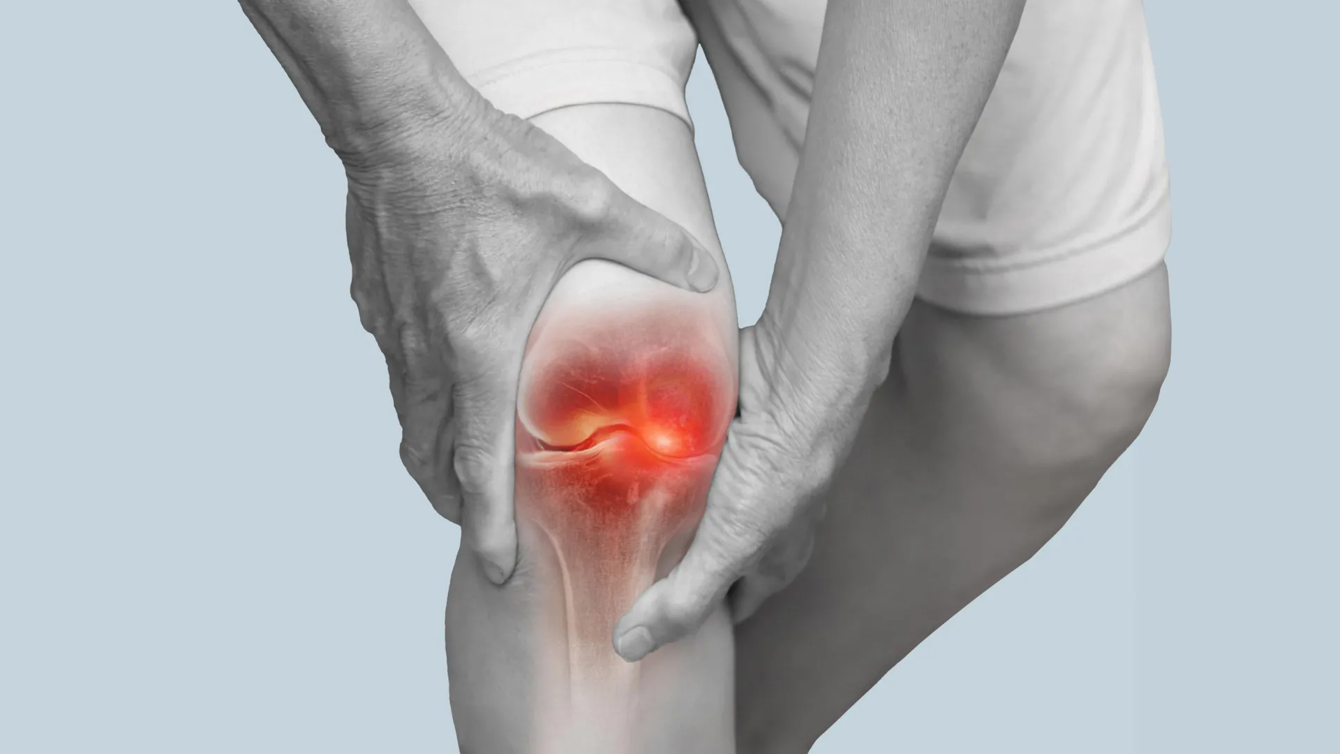
What if easing knee arthritis pain did not require another pill, an injection, or surgery, but simply changing how you walk?
A clinical trial published in The Lancet Rheumatology suggests that a small, carefully chosen adjustment to the angle of your feet could make a meaningful difference. After one year, participants who learned to walk with a personalized foot position reported significantly less knee pain than those receiving a placebo treatment. They also showed encouraging signs of slower deterioration in their knee cartilage.
The pain relief was comparable in magnitude to benefits reported for some commonly used medications. More importantly, the approach may help address something painkillers cannot: the excessive pressure on knee joints that contributes to osteoarthritis progression.
A Simple Walking Change Could Help Fight Knee Arthritis
Osteoarthritis affects roughly 22% of adults over age 40 and is one of the leading causes of disability. The condition gradually damages cartilage, the smooth, protective tissue that cushions the ends of bones and allows joints to move with minimal friction.
As cartilage deteriorates, everyday activities such as walking, climbing stairs, and getting out of a chair can become increasingly painful. Although exercise, physical therapy, weight management, and medications can help manage symptoms, there is currently no established treatment that reliably restores cartilage lost to osteoarthritis. People with advanced disease may eventually need joint replacement surgery.
Researchers from the University of Utah, New York University, and Stanford University have investigated a different strategy called gait retraining. Rather than focusing only on relieving pain, this approach changes the mechanics of walking to reduce pressure on vulnerable parts of the knee.
The findings, published in August 2025, came from the first randomized, placebo-controlled clinical trial to demonstrate the effectiveness of personalized gait retraining for knee osteoarthritis over a full year.
The research was co-led by Scott Uhlrich, an assistant professor of mechanical engineering at the University of Utah’s John and Marcia Price College of Engineering, with collaborators at Stanford and New York University. The work received support from federal research agencies, with the U.S. Department of Veterans Affairs listed as the trial’s funder.
“We’ve known that for people with osteoarthritis, higher loads in their knee accelerate progression, and that changing the foot angle can reduce knee load,” said Uhlrich, an assistant professor of mechanical engineering. “So the idea of a biomechanical intervention is not new, but there have not been randomized, placebo-controlled studies to show that they’re effective.”
Why the Angle of Your Feet Matters
The secret lies in how body weight moves through the knee with each step.
The knee has an inner section, called the medial compartment, and an outer section, known as the lateral compartment. During normal walking, the inner section typically carries more of the load. This helps explain why osteoarthritis affecting the inside of the knee is especially common.
For people with this type of arthritis, repeatedly placing excessive pressure on the already damaged area can accelerate deterioration.
Changing the direction in which the toes point during walking can redistribute some of that pressure. Turning the toes slightly inward or outward alters the forces acting on the knee, potentially reducing stress on the affected cartilage.
However, there is an important catch: the same adjustment does not work for everyone.
Some people benefit from turning their toes inward, while others respond better when their toes point farther outward. Making the wrong adjustment could fail to help or even increase pressure on the damaged joint.
“Previous trials prescribed the same intervention to all individuals, resulting in some individuals not reducing, or even increasing, their joint loading,” Uhlrich said. “We used a personalized approach to selecting each individual’s new walking pattern, which improved how much individuals could offload their knee and likely contributed to the positive effect on pain and cartilage that we saw.”
How Scientists Found the Right Walking Pattern
The researchers focused on people with mild to moderate osteoarthritis affecting the inner part of the knee.
At their first two laboratory visits, participants underwent MRI scans and walked on a specialized treadmill equipped with sensors that measured the forces generated during each step. Motion capture cameras recorded their movements in detail, allowing the researchers to analyze how different walking styles affected the knee.
Participants tested small changes in their foot position, turning their toes inward or outward by either 5° or 10°.
The scientists then identified which adjustment reduced pressure on the affected knee most effectively for each person.
Not everyone qualified. Some prospective participants were excluded because none of the tested adjustments successfully reduced their knee loading. Including such individuals in earlier studies may help explain why previous attempts at gait retraining produced inconsistent results.
After the initial assessments, 68 participants were randomly divided into two groups of 34.
One group received personalized instructions designed to minimize stress on the inner knee. The other received a sham treatment, meaning they went through the same training process but were instructed to maintain their natural foot angle.
This comparison allowed the researchers to separate the benefits of changing walking mechanics from improvements caused by expectations, attention from researchers, or participation in a structured walking program.
Just Six Weeks of Training Changed How They Walked
Both groups attended six weekly training sessions.
During these visits, participants walked on a treadmill while wearing a small device on their shin. The device provided biofeedback, delivering gentle vibrations whenever their foot moved outside the prescribed angle.
Biofeedback is a technique that uses immediate signals to help people recognize and gradually control movements they normally perform without thinking.
Over time, participants learned to maintain their assigned foot position without needing constant reminders.
After completing the six weeks of supervised training, they were encouraged to practice their walking technique for at least 20 minutes each day until it became a natural habit.
Follow-up assessments indicated that participants were remarkably consistent. On average, they maintained their prescribed foot angles to within approximately one degree.
That consistency is important because the potential benefits depend on continuing the altered walking pattern during ordinary daily activities, not just during laboratory sessions.
Arthritis Pain Improved, and Cartilage Showed Less Damage
One year after starting the program, participants returned for another MRI and reported how much knee pain they were experiencing.
The results showed a meaningful advantage for those who received personalized gait retraining.
On a pain scale from zero to 10, participants in the intervention group reported an average improvement of 2.5 points, compared with 1.3 points in the sham group.
In an exploratory analysis, approximately 91% of participants receiving personalized retraining experienced a clinically meaningful reduction in pain, compared with 66% in the comparison group.
The researchers also found that the intervention reduced a mechanical measure associated with pressure on the inner knee, confirming that the altered walking pattern had effects beyond participants’ perceptions of pain.
“The reported decrease in pain over the placebo group was somewhere between what you’d expect from an over-the-counter medication, like ibuprofen, and a narcotic, like OxyContin,” Uhlrich said. “With the MRIs, we also saw slower degradation of a marker of cartilage health in the intervention group, which was quite exciting.”
The MRI findings may be especially important.
Researchers used advanced imaging techniques capable of detecting subtle changes in cartilage composition before major structural damage becomes visible on conventional scans.
One measurement, known as T1rho, suggested that cartilage in the inner knee deteriorated more slowly among participants who changed their walking pattern. A second MRI measurement did not show a significant difference between the groups.
These findings do not establish that damaged cartilage was rebuilt or that arthritis progression can be permanently stopped. However, they raise the possibility that reducing mechanical stress could help preserve cartilage health over time.
The treatment also appeared generally well tolerated. No severe adverse events were reported, although two participants in the intervention group and one in the sham group withdrew because of increased knee pain.
More Recent Research Adds to the Evidence
Research published since the 2025 trial has provided further insight into how walking modifications might help people with knee osteoarthritis.
In a separate randomized trial published in Clinical Biomechanics in April 2026, researchers at the University of Bath investigated different types of biofeedback training in 50 people with knee osteoarthritis.
Participants completed a six-week program using different feedback strategies intended to improve walking mechanics.
All three groups, including the control group, experienced improvements in activity-related knee pain and physical function. However, only the group receiving feedback about its walking pattern achieved a lasting reduction in a key measure of knee loading.
That group’s knee loading measure fell by 7.6%, with the improvement maintained at a follow-up assessment one month later.
Interestingly, the investigators did not detect significant changes in foot angle in any of the groups. This suggests that other adjustments to walking mechanics can also influence the forces acting on the knee.
The findings reinforce an important lesson from the 2025 trial: simply encouraging people to walk differently is not necessarily enough. The type of movement being taught, and whether it successfully reduces harmful joint loading, appears to matter.
The 2026 study did not examine long-term cartilage deterioration, so it cannot establish whether these additional walking strategies slow osteoarthritis progression.
Could This Help People Avoid Knee Replacement?
For people living with knee osteoarthritis, the appeal of this approach extends beyond its measurable effects in the laboratory.
One trial participant described the freedom of adopting a treatment that did not require medication or an assistive device:
“I don’t have to take a drug or wear a device…it’s just a part of my body now that will be with me for the rest of my days, so that I’m thrilled with.”
Once a person learns the technique, it can potentially become part of ordinary daily movement without requiring continued use of specialized equipment.
That could be particularly valuable for people who develop arthritis relatively early in adulthood.
“Especially for people in their 30s, 40s, or 50s, osteoarthritis could mean decades of pain management before they’re recommended for a joint replacement,” Uhlrich said. “This intervention could help fill that large treatment gap.”
Whether gait retraining can actually delay or prevent knee replacement remains unknown. Longer studies would need to demonstrate that its benefits persist and translate into better joint health and fewer surgical procedures.
Still, the prospect of relieving pain while potentially slowing cartilage deterioration makes personalized walking adjustments an intriguing addition to the available treatment options.
Smart Shoes and Smartphone Cameras Could Make Treatment Easier
One major obstacle remains: delivering personalized gait retraining outside a specialized research laboratory.
The original trial relied on sophisticated motion capture equipment, pressure-sensitive treadmill measurements, MRI scans, and repeated in-person visits. Those resources are expensive and difficult to provide in ordinary physical therapy clinics.
Researchers are exploring ways to make the technology much simpler.
“We and others have developed technology that could be used to both personalize and deliver this intervention in a clinical setting using mobile sensors, like smartphone video and a ‘smart shoe’,” Uhlrich said.
Smartphone cameras could help analyze walking mechanics and estimate how different movements influence knee loading. Meanwhile, shoes fitted with motion sensors could provide feedback as patients practice walking at home or around their neighborhoods.
Work published in Machine Learning with Applications in September 2026 explored another possibility: using artificial intelligence to estimate knee loading from acceleration measurements collected at the foot.
In that proof of concept study, researchers analyzed walking data from 10 healthy adults and demonstrated that a machine learning system could track changes in estimated knee forces across different walking patterns.
The technology remains experimental and has not yet been validated as a treatment tool for people with knee osteoarthritis. Nevertheless, it illustrates how future rehabilitation systems might provide personalized guidance without requiring a room full of cameras and laboratory equipment.
A Promising Treatment, but Not a Do-It-Yourself Fix
Despite the encouraging results, researchers caution that personalized gait retraining is not yet ready for widespread clinical use.
The original study included only 68 participants, all of whom had mild to moderate osteoarthritis affecting the inner knee and demonstrated that at least one tested walking adjustment could reduce their joint loading. Its findings may not apply to people with advanced arthritis, other patterns of joint damage, or those whose knees respond differently.
Importantly, turning the toes inward or outward without professional assessment is not the same as receiving personalized gait retraining. An adjustment that helps one person might increase stress on another person’s knee.
Larger trials, longer follow-up periods, and practical methods for delivering the treatment will be needed before clinicians can routinely prescribe it.
For now, the research points to an intriguing possibility: the way people walk may influence not only how much their knees hurt today, but potentially how well their cartilage holds up in the years ahead.

Scientists at the U.S.Department of Energy’s (DOE) Princeton Plasma Physics Laboratory (PPPL) have identified a potentially much more efficient way to achieve fusion ignition, a major milestone in the pursuit of practical fusion energy. Their calculations suggest that changing the order in which plasma is heated and compressed could dramatically reduce the energy needed to reach a self-sustaining fusion reaction.
For more than seven decades, fusion researchers have relied on a mathematical rule to determine whether plasma can remain sufficiently hot and dense, for a long enough period, to sustain fusion without additional external heating. Known as the Lawson criterion, this equation defines the conditions required for ignition. However, it does not explain the most efficient way to achieve them.
Now, PPPL physicists Luis Delgado-Aparicio, Masayuki Ono, and Jonathan Menard have developed a more comprehensive version of the criterion. By incorporating four additional factors that influence fusion performance, they have mapped a promising route to ignition that could require substantially less energy than alternative approaches.
Their findings were published in the journal Physical Review Letters.
The research focuses on plasma, often called the fourth state of matter. Plasma forms when a gas becomes so energetic that its particles carry electrical charges. In fusion reactors, this extremely hot material contains the atomic nuclei that researchers hope to combine, releasing energy through nuclear fusion.
The ultimate goal is to produce a burning plasma, in which fusion reactions generate enough heat to sustain the process without continued external heating. Reaching this state efficiently remains one of the most important challenges in fusion energy research.
Scientists Find a Shortcut to Fusion Ignition
Delgado-Aparicio compares the discovery to finding a more efficient route through a mountain range.
Imagine that the energy requirements for fusion ignition form a landscape dominated by a towering mountain. The destination lies on the other side, but there are several possible routes to reach it.
Many proposed fusion strategies effectively attempt to climb directly over the mountain. They begin by increasing the density of the plasma and then supplying the enormous amount of heat needed to reach ignition.
The new calculations suggest that a different sequence could be considerably more efficient. Instead of increasing density first, researchers could heat the plasma before making it denser. This alternative route avoids the highest energy requirements while still reaching the desired conditions.
“A lot of companies want to climb the mountain head-on and spend enormous energy to get there,” Delgado-Aparicio said. “Go around the peak instead. You reach the same place in a much smarter way, and you use far less energy.”
The Hidden Mountain Pass in Fusion Physics
The researchers identified a particularly important region of this mathematical landscape called the Cordey saddle.
In geographical terms, a saddle is a low passage between higher elevations. In fusion physics, the Cordey saddle represents a relatively accessible point along the boundary separating plasma that still needs outside heating from plasma capable of sustaining its own fusion reactions.
To evaluate different routes, the scientists used a measurement called Q, which compares the power produced by fusion reactions with the external heating power supplied to the plasma.
For example, a Q value of 5 means the fusion reactions produce five times as much power as the external heating system delivers.
In an idealized plasma containing only pure fusion fuel, the Cordey saddle occurs at approximately Q = 5. However, actual fusion systems are much more complicated. Contaminants within the plasma and extremely strong magnetic fields can shift the location of this passage, increasing the Q value needed to reach it.
Understanding how these conditions change is essential for determining whether a proposed fusion reactor can realistically achieve ignition.
A More Realistic Roadmap for Future Fusion Reactors
The findings could influence the design of two major types of magnetic fusion devices: tokamaks and stellarators.
Both approaches use powerful magnetic fields to confine plasma while it is heated to the extraordinary temperatures needed for fusion. Tokamaks, which have a characteristic doughnut shape, are among the leading technologies being investigated for future fusion power plants.
Research institutions and private companies are pursuing several different ways to generate electricity from fusion and eventually deliver that power to the electrical grid. Identifying an efficient route to ignition could help engineers design more practical systems while avoiding costly mistakes.
For PPPL, this work is part of a broader effort to address the scientific and technological challenges that must be overcome before commercial fusion power becomes possible, including the development of future fusion pilot plants.
One of the study’s major advances is that it evaluates several important physical processes together rather than examining them separately, as much previous research has done.
The researchers incorporated four additional factors that can influence whether a plasma reaches and maintains ignition:
Accounting for these effects produces a more realistic picture of the conditions future reactors must achieve.
“When you leave these effects out, you say the design will work fine,” Ono said. “When you put them in, the picture changes, and it becomes quite important.”
According to Ono, the goal is to establish a more reliable, widely applicable model that scientists can use to evaluate fusion reactor designs before construction begins.
Treating the Lawson criterion as a single unchanging threshold overlooks physical processes that can substantially alter the requirements for ignition. Including those effects early in the design process could prevent expensive problems later.
“Fusion experiments cost a great deal of money, and you do not want to make mistakes you could have caught beforehand,” he said.
Tiny Amounts of Tungsten Could Make Fusion Much Harder
One particularly striking finding concerns tungsten, an extremely heat-resistant metal selected for the interior walls of more than a dozen next-generation fusion machines.
Tungsten is attractive because fusion reactors must withstand conditions involving plasma temperatures greater than those found at the center of the sun. However, the material also presents a significant challenge if even tiny quantities enter the plasma.
The team’s calculations show that tungsten contamination at a concentration of just one part in 10,000 can approximately double the pressure required to achieve fusion ignition.
That is a substantial increase caused by an exceptionally small amount of material.
The calculations were initially developed in two dimensions. When the researchers extended their analysis to three dimensions, they found that the pressure needed for ignition could exceed the limits at which the plasma remains stable.
This suggests that seemingly minor contamination could have major consequences for the feasibility of certain fusion reactor designs.
How Energy Losses Could Help Stabilize Fusion
Surprisingly, some of the processes that make ignition more difficult may also help fusion reactors operate more safely and consistently.
Scientists have long investigated the possibility of thermal “runaway” instability. This occurs when heat from fusion reactions increases the reaction rate, producing still more heat and potentially creating a self-reinforcing cycle.
The researchers found that energy losses within the plasma can naturally counteract this effect.
Although those losses increase the difficulty of reaching ignition, they also provide a balancing influence that could allow a burning plasma to maintain a stable operating state without constant adjustments.
This balance could be valuable for future reactor designs.
(Unlike a fission power plant, a fusion system holds only seconds of fuel at a time, so losing control means the plasma cools and the burn stops rather than anything worse.)
Liquid Lithium and Advanced Fuel Could Improve Fusion Efficiency
The study also identifies technologies that might help overcome some of the obstacles revealed by the calculations.
One possibility involves coating the inside of fusion reactors with liquid lithium, a technique PPPL researchers have investigated for years.
These coatings could help prevent tungsten from entering the plasma while reducing heat losses, making it easier to maintain the extreme conditions necessary for fusion.
Another promising approach uses spin-polarized fuel. This involves preparing the atomic nuclei so that their spins are aligned before fusion occurs, which can increase the fusion reaction rate.
Together, these strategies could improve the performance of future reactors and make the path to ignition more achievable.
What Comes Next for the New Fusion Strategy?
Despite the promising findings, the proposed heat-first approach has not yet been demonstrated experimentally.
The research is theoretical and relies on mathematical calculations rather than direct measurements. Existing fusion experiments have not reached the temperatures associated with the Cordey saddle, preventing scientists from testing the predicted route under those conditions.
The PPPL team plans to investigate the approach further through digital experiments, using computer-based simulations to explore whether heating plasma before increasing its density can deliver the expected advantages.
If future studies confirm the calculations, the findings could provide an important foundation for designing more efficient fusion systems and help researchers make better decisions about reactor construction.
“While more study is needed, we are excited by these results, and they suggest a clear path forward for future research in this area,” said Menard.
The work was supported by the DOE Office of Science, Office of Fusion Energy Sciences, under contract DE-AC02-09CH11466. Additional support came through a 2015 Fusion Energy Sciences Early Career Award, a 2018 Diagnostic Innovation and Development award, and the 2021 and 2025 Long-Pulse Tokamak Research Programs.

Astronomers have discovered an unusual star system about 300 light-years from Earth where a small star is gradually consuming a nearby brown dwarf. Instead of swallowing its companion in one dramatic event, the star is steadily pulling material from it, creating a cosmic feeding process that could continue for billions of years.
Most planets, including Earth, travel around their stars in relatively stable orbits. But these peaceful arrangements can sometimes end violently. If a planet ventures too close to its host star, the star’s gravity can drag it inward and eventually engulf it.
Scientists have observed many planetary systems with stable orbits, along with a small number of stars caught rapidly swallowing their planetary companions. Now, researchers have identified something that falls between those two extremes.
In a study published October 5 in Nature Astronomy, an international team led by scientists at MIT describes a star that is slowly feeding on a brown dwarf, an object more massive than a typical planet but not massive enough to sustain the hydrogen fusion that powers ordinary stars.
The system, designated ZTF J0440+2325, lies within our Milky Way galaxy approximately 300 light-years away. It is the first observed example of a low-mass star steadily drawing material from another low-mass object.
Even more remarkably, the transfer of material appears to be happening so slowly that the star could continue feeding on its companion for hundreds of thousands of years, potentially even billions.
“When we think of stars interacting with planets or brown dwarfs, the picture is always that the star eventually swallows the other thing,” says Kevin Burdge, assistant professor of physics at MIT. “This is what will happen to the Earth when the sun becomes a red giant. But here, we’ve found an alternative: Instead of swallowing the thing up, the star can gradually eat it, for billions of years.”
The MIT research team also includes Aaron Householder, Kaitlyn Shin, Saul Rappaport, Joheen Chakraborty, and Emma Chickles. Additional collaborators contributed from Caltech, the University of Hawaii, the Instituto de Astrofísica de Canarias and Universidad de La Laguna in Spain, and the Harvard-Smithsonian Center for Astrophysics.
A Mysterious Signal That Puzzled Astronomers for Years
The first clue to this unusual stellar relationship came from the Zwicky Transient Facility (ZTF), an astronomical survey that searches for objects whose brightness changes over time.
Operating with a telescope at Palomar Observatory in California, ZTF repeatedly photographs large sections of the night sky. Its sensitive camera can detect sudden or unusual changes in light that may indicate powerful cosmic events, including exploding stars called supernovae, gamma-ray bursts, and collisions between neutron stars.
Several years ago, while examining observations collected by ZTF, Burdge noticed a particularly strange pattern.
Astronomers study changes in an object’s brightness using a graph called a light curve. For some supernovae, these graphs form a rounded, bell-like shape as the exploding star grows brighter and then gradually fades.
The signal Burdge encountered looked completely different. Instead of a smooth rise and fall, the brightness repeatedly traced something resembling a triangle.
“I remember first looking at this and thinking, stars don’t make triangular waveforms like this,” he recalls.
At the time, Burdge and his colleagues were investigating another unusual signal that turned out to originate from a system known as a “black widow binary.”
These systems contain a neutron star, the extraordinarily dense remnant of a massive star, that spins rapidly while stripping material from a much smaller companion. They are named after black widow spiders because of the way the neutron star gradually destroys its partner.
Burdge initially considered whether the repeating triangular signal might come from another black widow system. However, the observations did not match what astronomers normally expect.
In a typical black widow binary, the strong contrast between the masses of the two objects produces a distinctive motion. The smaller companion moves rapidly around the much heavier neutron star, creating detectable changes in the light reaching Earth.
But the mysterious triangular signal showed no evidence of the same pronounced motion.
“We weren’t seeing that whipping back and forth here,” Burdge says. “It didn’t make any sense. We couldn’t explain what this was.”
That discrepancy led the researchers to consider another possibility. Perhaps the system did not contain one extremely massive object paired with a much lighter companion. Instead, both objects might have relatively small masses, allowing them to orbit each other with much less dramatic motion.
“If you have less mass in the system overall, things can gently orbit each other without whipping back and forth,” Burdge says. “That was the idea. But we never had any proof. And this weird triangle just sat for years.”
A Brown Dwarf That Orbits Its Star Every 87 Minutes
Eventually, Burdge and Aaron Householder, a graduate student in MIT’s Department of Earth, Atmospheric, and Planetary Sciences, returned to the unexplained signal.
Using the original ZTF observations, they identified its source as an object roughly 300 light-years away in the Milky Way. They then directed several telescopes toward the system, ZTF J0440+2325, to investigate its properties in greater detail.
A key measurement involved determining how much the system’s objects move in response to their mutual gravitational attraction. Although the researchers detected some wobbling, it was far smaller than the motion typically associated with black widow binaries.
“That was the real clincher for this system,” Householder says. “When we measured that wobble, we found we were not seeing a black widow. This was a low-mass star that’s orbited by a brown dwarf. The wobble was too small in amplitude to be anything else.”
The observations revealed two unusually compact objects orbiting at an extremely close distance.
The central star has approximately 85 times the mass of Jupiter, while the brown dwarf weighs about 25 Jupiter masses. For comparison, the Sun is more than 1,000 times as massive as Jupiter.
The brown dwarf completes a full revolution around its host star in just 87 minutes. Its entire orbit is so small that it would fit within the diameter of the Sun.
That extraordinarily close arrangement suggested that gravity might be doing more than simply keeping the objects in orbit. The researchers suspected that the star could be actively pulling matter away from the brown dwarf.
Scientists Discover a New Form of Stellar Cannibalism
When one astronomical object draws gas or other material from another, astronomers call the process accretion.
Accretion is especially familiar in systems involving black holes and neutron stars. Although these objects can be extremely massive, their physical dimensions are relatively small. Material falling toward a black hole, for example, often forms a swirling disk around it before moving inward.
But ZTF J0440+2325 appears to work differently.
Rather than a black hole or neutron star collecting matter, an ordinary low-mass star is receiving material from a nearby brown dwarf. Because the star has a much larger physical surface than a compact object such as a black hole, the incoming material can strike it directly.
“The difference here is, the thing absorbing matter is not a tiny black hole but a star, which is relatively big in size,” Burdge explains. “So matter just pummels directly onto the surface, at very high speeds, like an asteroid hitting the Moon.”
To investigate whether this was actually happening, the researchers created computer simulations of the system.
They modeled particles originating from the brown dwarf and calculated how those particles would move under the gravitational influence of both objects. The simulations incorporated the measured properties of the star and its companion, along with the physical equations governing orbital motion.
The results supported the team’s suspicion. Material leaving the brown dwarf followed trajectories that carried it directly into the star.
“When we track those test particles, we see they indeed fall right onto the surface of the star,” Householder says. “This is the first time we’ve caught a low-mass star actively accreting from another low-mass object.”
A Cosmic Feast That Could Continue for Billions of Years
The team also estimated how quickly the brown dwarf is losing material.
According to their calculations, the star is consuming approximately one hundred-thousandth of Earth’s mass every year.
That amount may sound insignificant, but it represents an enormous quantity of material. The researchers compare it to approximately 40 million dump trucks’ worth of matter, or about 1.3 trillion one-pound burritos every second.
Despite those staggering figures, the brown dwarf is so massive that this material loss represents only a tiny portion of its total mass.
In astronomical terms, the feeding process is remarkably gradual.
Based on the properties of the system and the estimated rate of mass transfer, researchers believe the star could continue drawing matter from its companion for billions of years.
Instead of a sudden, catastrophic collision that destroys the smaller object, the star appears to be sustaining a relatively steady flow of material from its neighbor.
That realization also helped explain the strange triangular light pattern that first attracted Burdge’s attention.
As matter streams from the brown dwarf toward the star, it strikes the stellar surface at high speeds, generating an intensely heated region.
This bright hotspot acts like a persistent fireball on the star.
As the brown dwarf moves through its 87-minute orbit, the hotspot repeatedly rotates into and out of the observer’s view. Those changes in visibility produce the distinctive pattern of brightening and dimming that the astronomers had struggled to understand.
“It’s like you’ve got this continuous fireball onto one of the objects, and as one orbits the other, that hotspot comes in and out of view, and the peak of the triangle signal is when you’re looking right at the fireball,” Burdge explains.
A Strange Star System Opens a New Window Into Planetary Evolution
With the triangular signal finally explained, the researchers are now interested in identifying other stellar systems that behave in similar ways.
Until now, observations of close interactions between stars and their planetary companions have largely emphasized either stable orbits or relatively rapid engulfment events.
ZTF J0440+2325 demonstrates that another possibility exists. Under the right conditions, a small star can steadily extract matter from a nearby brown dwarf without immediately destroying it.
Finding additional examples could help astronomers determine how common this process is, how long it can persist, and what ultimately happens to objects trapped in such extraordinarily close relationships.
The discovery may also provide new insights into the evolution of planets and brown dwarfs that orbit near their host stars.
“It’s inspiring a lot of new searches on our part,” Householder says. “I think we’re going to learn a lot about a different kind of way that planets and brown dwarfs interact with their host stars.”
This research received partial support from the National Science Foundation.
