Lab grown human spinal cord heals after injury in major breakthrough

Scientists at Northwestern University have created the most sophisticated lab grown model yet for studying human spinal cord injury.

In the new research, the team worked with human spinal cord organoids — miniature organs derived from stem cells — to recreate different forms of spinal cord trauma and evaluate a promising regenerative treatment.

For the first time, researchers showed that these human spinal cord organoids can faithfully reproduce the major biological consequences of spinal cord injury. The model displayed cell death, inflammation, and glial scarring, which is a thick buildup of scar tissue that forms a physical and chemical barrier preventing nerve repair.

When the damaged organoids were treated with “dancing molecules” — a therapy that restored movement and repaired tissue in a previous animal study — the results were dramatic. The injured tissue produced substantial neurite outgrowth, meaning the long extensions that allow neurons to communicate began growing again. Scar like tissue was greatly reduced. The findings add support to the idea that this therapy, which recently received Orphan Drug Designation from the U.S. Food and Drug Administration (FDA), could improve recovery for people with spinal cord injuries.

The study was published on Feb. 11 in Nature Biomedical Engineering.

“One of the most exciting aspects of organoids is that we can use them to test new therapies in human tissue,” said Northwestern’s Samuel I. Stupp, the study’s senior author and inventor of dancing molecules. “Short of a clinical trial, it’s the only way you can achieve this objective. We decided to develop two different injury models in a human spinal cord organoid and test our therapy to see if the results resembled what we previously saw in the animal model. After applying our therapy, the glial scar faded significantly to become barely detectable, and we saw neurites growing, resembling the axon regeneration we saw in animals. This is validation that our therapy has a good chance of working in humans.”

Stupp is a leader in regenerative materials science and holds the title of Board of Trustees Professor of Materials Science and Engineering, Chemistry, Medicine and Biomedical Engineering at Northwestern. He has appointments in the McCormick School of Engineering, Weinberg College of Arts and Sciences and Feinberg School of Medicine, and directs the Center for Regenerative Nanomedicine (CRN). The paper’s first author is Nozomu Takata, a research assistant professor of medicine at Feinberg and member of CRN.

Why Human Organoids Matter

Organoids are grown from induced pluripotent stem cells in the laboratory. Although they are simplified versions of full organs, they closely resemble real tissue in structure, cellular diversity, and function. Because of this, organoids are powerful tools for studying disease, testing treatments, and exploring how organs develop. They also allow researchers to move faster and at lower cost compared to animal experiments or human clinical trials.

While other groups have produced spinal cord organoids to study basic biology, this model represents a major advance for injury research. The organoids measured several millimeters across and were mature enough to sustain and model traumatic damage.

Over several months, the team guided stem cells to form complex spinal cord tissue containing neurons and astrocytes. They also became the first to incorporate microglia — immune cells found in the central nervous system — to better replicate the inflammatory response that follows spinal cord injury.

“It’s kind of a pseudo-organ,” Stupp said. “We were the first to introduce microglia into a human spinal cord organoid, so that was a huge accomplishment. It means that our organoid has all the chemicals that the resident immune system produces in response to an injury. That makes it a more realistic, accurate model of spinal cord injury.”

What Are Dancing Molecules

Once the spinal cord organoids were fully developed, the researchers turned their attention to testing injury and treatment. First introduced in 2021, the dancing molecules therapy uses controlled molecular motion to repair tissue and potentially reverse paralysis after traumatic spinal cord injury. It belongs to a broader class of supramolecular therapeutic peptides (STPs), which rely on large assemblies of 100,000 or more molecules to activate cell receptors and stimulate the body’s natural repair signals. (The concept of supramolecular therapies also is used in current GLP-1 drugs for weight loss and diabetes, an area that Stupp’s lab investigated nearly 15 years ago.)

The therapy is delivered as a liquid injection that quickly forms a web of nanofibers resembling the spinal cord’s extracellular matrix. By adjusting how dynamically the molecules move within this structure, researchers improved how effectively they interact with constantly shifting cell receptors.

“Given that cells themselves and their receptors are in constant motion, you can imagine that molecules moving more rapidly would encounter these receptors more often,” Stupp said in 2021. “If the molecules are sluggish and not as ‘social,’ they may never come into contact with the cells.”

In previous animal experiments, a single injection given 24 hours after a severe injury enabled mice to walk again within four weeks. Formulations with faster molecular motion performed better than slower versions, suggesting that increased movement enhances bioactivity and cellular signaling.

Simulating Spinal Cord Trauma

To test the therapy, the researchers created two common types of spinal cord injury in the organoids. Some were cut with a scalpel to mimic a laceration similar to a surgical wound. Others were subjected to a compressive contusion injury, comparable to trauma from a serious car crash or fall.

Both types of injury led to cell death and the formation of glial scars — just as occurs in real spinal cord injury.

“We could distinguish between the astrocytes that are a part of normal tissue and the astrocytes in the glial scar, which are large and very densely packed,” Stupp said. “We also detected the production of chondroitin sulfate proteoglycans, which are molecules in the nervous system that respond to injury and disease.”

After treatment with dancing molecules, the gelled nanofiber scaffold reduced inflammation, shrank glial scarring, stimulated neurite extension, and encouraged neurons to grow in organized patterns.

Neurites include axons, which are often severed in spinal cord injuries. When axons are cut, communication between neurons is disrupted, leading to paralysis and loss of sensation below the injury site. Promoting neurite regrowth could reconnect these pathways and help restore function.

The Role of Molecular Motion

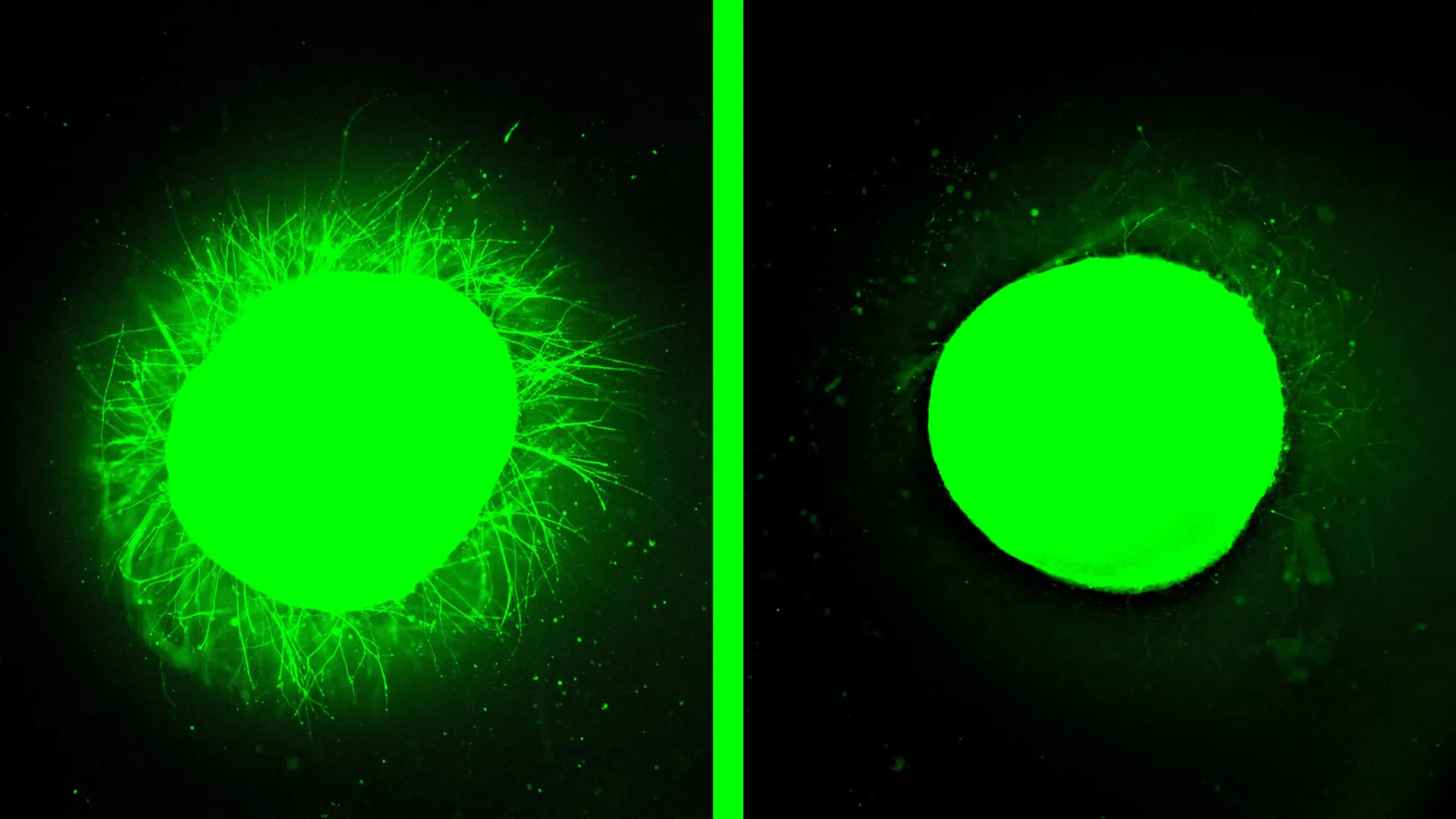
Stupp credits the therapy’s effectiveness to supramolecular motion, meaning the ability of the molecules to move rapidly and even briefly detach from the nanofiber network. Experiments on healthy organoids reinforced this idea.

“Before we even developed the injury model, we tested the therapy on a healthy organoid,” he said. “The dancing molecules spun out all these long neurites on the surface of the organoid but, when we used molecules that had less or no motion, we saw nothing. This difference was very vivid.”

Looking ahead, the team plans to engineer even more advanced organoids to refine their models. They also intend to develop versions that replicate chronic, long standing injuries, which typically involve thicker and more persistent scar tissue. With further development, Stupp said these miniature spinal cords could contribute to personalized medicine by generating implantable tissue from a patient’s own stem cells, reducing the risk of immune rejection.

The study, “Injury and therapy in a human spinal cord organoid,” was supported by the Center for Regenerative Nanomedicine at Northwestern University and a gift from the John Potocsnak Family for spinal cord injury research.

Share Button

Brain inflammation may be driving compulsive behavior

For years, scientists have believed that compulsive behaviors happen when people become trapped in a “habit loop” that overrides self-control. But new research in rats from the University of Technology Sydney (UTS) suggests the story may be more complicated.

Compulsive behaviors appear in a range of mental health conditions, including obsessive-compulsive disorder, substance use disorders, and gambling disorder. In these conditions, people continue repeating certain actions even when they lead to harmful consequences. Millions of people worldwide are affected.

How Habits and Self-Control Normally Work

Senior author Dr. Laura Bradfield, a behavioral neuroscientist, explained that habits serve an important purpose. They allow us to run on autopilot during routine tasks like brushing our teeth or driving along a familiar road, freeing up mental energy for other thoughts.

“However, if we are driving and a child steps onto the road, then we suddenly become aware of our surroundings and focus on what we are doing. This involves taking back conscious control, thinking about possible outcomes and adjusting our behavior,” said Dr. Bradfield.

In compulsive behaviors such as repeated handwashing or playing poker machines, the prevailing theory has been that these actions become deeply ingrained habits. According to this view, the behavior runs automatically, making it hard for people to regain cognitive control.

“Brain imaging studies show it’s common for people with compulsive disorders to have inflammation in the striatum, a brain region involved in choosing actions, so we decided to test whether inducing inflammation in this region in rats would increase habitual behavior.”

Brain Inflammation and Decision-Making

The study was led by Dr. Arvie Abiero during his PhD research at UTS and was recently published in Neuropsychopharmacology. The researchers examined how rats learn behaviors and how they regulate their actions. When inflammation was triggered in the striatum, the results were unexpected. Instead of becoming more automatic or habit-driven, the rats showed more deliberate and effortful decision-making.

“Surprisingly, the animals became more goal-directed and continued to adjust their behavior based on outcomes, even in situations where habits would normally take over,” said Dr. Bradfield.

The Role of Astrocytes in Compulsive Behavior

The team traced these changes to astrocytes, star-shaped cells in the brain that support neurons. When inflammation occurred, astrocytes multiplied and disrupted nearby neural circuits that control movement and decision-making.

These findings could have important implications for psychologists, psychiatrists, patients, and caregivers who work with compulsive disorders. Rather than reflecting a loss of control due to runaway habits, some compulsive behaviors may result from excessive, though misdirected, deliberate control.

The researchers suggest that medications aimed at astrocytes or treatments that reduce neuroinflammation may provide new therapeutic options. Broader anti-inflammatory strategies, such as regular exercise or improved sleep, could also play a role.

“There’s a lot of compulsive behavior that doesn’t fit neatly into the habit hypothesis. If someone is continually washing their hands because they are worried about germs, they are not doing this without thinking, they are consciously choosing to make that effort,” said Dr. Bradfield.

“Our findings offer a new explanation for these behaviors, which goes against the accepted view. Based on this, it’s possible that new treatments and interventions can be developed that more effectively treat these diseases and disorders,” she said.

Share Button

Measles outbreak could see unvaccinated pupils excluded from schools in north London

Children identified as close contacts of people with the disease could be excluded for three weeks

Share Button

Women calling for greater support with menopause

Women who experienced challenges during the menopause set up businesses to help others.

Share Button

‘Fast-spreading’ measles outbreak hits several schools in London

A number of children have been hospitalised as officials urge families to check they are up to date with their vaccines.

Share Button

Large study finds no link between mRNA COVID vaccine in pregnancy and autism

Receiving an mRNA COVID-19 vaccine shortly before or at any point during pregnancy is not linked to autism or other developmental disorders in children, according to research presented at the Society for Maternal-Fetal Medicine (SMFM) 2026 Pregnancy Meeting. The findings provide new data on the long term health of children whose mothers were vaccinated around the time of pregnancy.

In the United States, two types of COVID-19 vaccines are recommended: the messenger ribonucleic acid (mRNA) vaccine and a protein subunit vaccine. Both are considered safe during all stages of pregnancy and are recommended to help safeguard both maternal and infant health.

Study of 434 Toddlers

The investigation was conducted by researchers within the Maternal-Fetal Medicine Units Network. The team evaluated 434 children between 18 months and 30 months of age for signs of autism and other developmental concerns.

The study was prospective, multi-center, and observational, and took place between May 2024 and March 2025. Half of the children (217) were born to mothers who received at least one dose of an mRNA COVID-19 vaccine either during pregnancy or within 30 days before becoming pregnant. The remaining 217 children were born to mothers who did not receive an mRNA vaccine during or within 30 days prior to pregnancy.

“Neurodevelopment outcomes in children born to mothers who received the COVID-19 vaccine during or shortly before pregnancy did not differ from those born to mothers who did not receive the vaccine,” said senior researcher George R. Saade, MD, Professor and Chair of Obstetrics and Gynecology, and Associate Dean for Women’s Health, at Macon & Joan Brock Virginia Health Sciences at Old Dominion University in Norfolk, VA.

How Researchers Compared Developmental Outcomes

To make the comparison as accurate as possible, vaccinated mothers were paired with unvaccinated mothers based on where they delivered (hospital, birth center, etc.), the date of delivery, insurance status, and race. Certain pregnancies were excluded from both groups, including those that ended before 37 weeks, involved multiple babies, or resulted in a child with a major congenital malformation.

When the children reached 1 ½ — 2 ½ years of age, researchers assessed their development using the Ages and Stages Questionnaire Version 3. This screening tool measures progress in five areas: communication, gross motor skills, fine motor skills, problem solving, and personal social interaction. The team also reviewed results from the Child Behavior Checklist, Modified Checklist for Autism in Toddlers, and the Early Childhood Behavior Questionnaire to further evaluate behavioral and developmental patterns.

“This study, conducted through a rigorous scientific process in an NIH clinical trials network, demonstrates reassuring findings regarding the long-term health of children whose mothers received COVID-19 vaccination during pregnancy,” said Brenna L. Hughes, MD, MSc, Edwin Crowell Hamblen Distinguished Professor of Reproductive Biology and Family Planning and Interim Chair of the Department of Obstetrics and Gynecology at Duke University in Raleigh, NC.

Funding and Disclosure

The study was funded by the Eunice Kennedy Shriver National Institute of Child Health and Human Development. The authors noted that the conclusions presented are their own and do not necessarily reflect the official views of the National Institutes of Health.

Oral abstract #8 “Association between SARS-CoV-2 vaccine in pregnancy and child neurodevelopment at 18-30 months” will be published in the February 2026 issue of PREGNANCY, the official peer-reviewed medical journal of the Society for Maternal-Fetal Medicine.   

Share Button

AI uncovers the hidden genetic control centers driving Alzheimer’s

A team led by Min Zhang and Dabao Zhang at the University of California, Irvine’s Joe C. Wen School of Population & Public Health has developed the most comprehensive maps yet of how genes directly influence one another in brain cells affected by Alzheimer’s disease. These maps go beyond identifying gene links. They reveal which genes are actively controlling others across different cell types in the brain.

To accomplish this, the researchers created a machine learning platform called SIGNET. Unlike traditional tools that only detect genes that appear to move together, SIGNET is designed to uncover true cause-and-effect relationships. Using this approach, the team identified important biological pathways that may contribute to memory loss and the gradual breakdown of brain tissue.

The findings were published in Alzheimer’s & Dementia: The Journal of the Alzheimer’s Association. The study also highlights newly identified genes that could become promising targets for future treatments. Funding support came in part from the National Institute on Aging and the National Cancer Institute.

Why Understanding Gene Control Matters in Alzheimer’s

Alzheimer’s disease is the leading cause of dementia and is expected to affect nearly 14 million Americans by 2060. Although scientists have linked several genes to the disease, including APOE and APP, they still do not fully understand how these genes interfere with normal brain function.

“Different types of brain cells play distinct roles in Alzheimer’s disease, but how they interact at the molecular level has remained unclear,” said Min Zhang, co-corresponding author and professor of epidemiology and biostatistics. “Our work provides cell type-specific maps of gene regulation in the Alzheimer’s brain, shifting the field from observing correlations to uncovering the causal mechanisms that actively drive disease progression.”

How SIGNET Reveals Cause and Effect Between Genes

To build these detailed maps, the team analyzed single-cell molecular data from brain samples donated by 272 participants enrolled in long-term aging studies known as the Religious Orders Study and the Rush Memory and Aging Project. SIGNET was designed as a scalable, high-performance computing system that combines single-cell RNA sequencing with whole-genome sequencing data. This integration allowed the researchers to detect cause-and-effect relationships among genes across the entire genome.

Using this method, they constructed causal gene regulatory networks for six major brain cell types. This made it possible to determine which genes are likely directing the activity of others, something conventional correlation-based methods cannot reliably accomplish.

“Most gene-mapping tools can show which genes move together, but they can’t tell which genes are actually driving the changes,” said Dabao Zhang, co-corresponding author and professor of epidemiology and biostatistics. “Some methods also make unrealistic assumptions, such as ignoring feedback loops between genes. Our approach takes advantage of information encoded in DNA to enable the identification of true cause-and-effect relationships between genes in the brain.”

Major Genetic Rewiring in Excitatory Neurons

The researchers found that the most significant gene disruptions occur in excitatory neurons — the nerve cells that send activating signals — where nearly 6,000 cause-and-effect interactions revealed extensive genetic rewiring as Alzheimer’s progresses.

The team also identified hundreds of “hub genes” that function as central regulators, influencing many other genes and likely playing an important role in harmful changes in the brain. These hub genes could become valuable targets for earlier diagnosis and future therapies. The study further uncovered new regulatory roles for well-known genes such as APP, which was shown to strongly control other genes in inhibitory neurons.

To strengthen their conclusions, the researchers validated their findings using an independent set of human brain samples. This additional confirmation increases confidence that the observed gene relationships reflect genuine biological mechanisms involved in Alzheimer’s disease.

Beyond Alzheimer’s, SIGNET may also be applied to the study of other complex diseases, including cancer, autoimmune disorders and mental health conditions.

Share Button

Stop eating 3 hours before bed to improve heart health

Researchers at Northwestern Medicine explored whether timing an overnight fast to match a person’s natural sleep wake cycle could improve heart and metabolic health. The circadian rhythm plays a central role in regulating cardiovascular and metabolic function. Importantly, participants did not reduce calories. The focus was entirely on adjusting when they ate.

The study found that middle age and older adults at elevated risk for cardiometabolic disease benefited from extending their overnight fasting window by roughly two hours. They also avoided food and dimmed lights for three hours before going to sleep. These changes led to measurable improvements in heart and metabolic markers during sleep and throughout the following day.

“Timing our fasting window to work with the body’s natural wake-sleep rhythms can improve the coordination between the heart, metabolism and sleep, all of which work together to protect cardiovascular health,” said first author Dr. Daniela Grimaldi, research associate professor of neurology in the division of sleep medicine at Northwestern University Feinberg School of Medicine.

The findings were published Feb. 12 in Arteriosclerosis, Thrombosis, and Vascular Biology, a journal of the American Heart Association.

“It’s not only how much and what you eat, but also when you eat relative to sleep that is important for the physiological benefits of time-restricted eating,” said corresponding author Dr. Phyllis Zee, director of the Center for Circadian and Sleep Medicine and chief of sleep medicine in the department of neurology at Feinberg.

Why Cardiometabolic Health Matters

Earlier data show that only 6.8% of U.S. adults had optimal cardiometabolic health in 2017 to 2018. Poor cardiometabolic health raises the risk of chronic conditions such as type 2 diabetes, non-alcoholic fatty liver disease, and cardiovascular disease.

Time-restricted eating has grown in popularity because studies suggest it can improve cardiometabolic markers and sometimes match the benefits of traditional calorie restricted diets. However, most research has concentrated on how long people fast rather than how well that fasting window aligns with sleep timing, which is crucial for metabolic regulation.

With nearly 90% adherence in this trial, the researchers believe anchoring time-restricted eating to the sleep period may be a realistic and accessible non-pharmacological approach, especially for middle age and older adults who face higher cardiometabolic risk.

The team plans to refine this protocol and expand testing in larger multi-center trials.

Blood Pressure, Heart Rate, and Blood Sugar Improvements

The 7.5 week study compared individuals who stopped eating at least three hours before bedtime with those who maintained their usual eating habits. Those who adjusted their timing experienced several meaningful changes.

Nighttime blood pressure decreased by 3.5%, and heart rate dropped by 5%. These shifts reflected a healthier daily pattern, with heart rate and blood pressure rising during daytime activity and falling at night during rest. A stronger day night rhythm is associated with better cardiovascular health.

Participants also demonstrated improved daytime blood sugar control. When given glucose, their pancreas responded more effectively, suggesting improved insulin release and steadier blood sugar levels.

The trial included 39 overweight/obese adults (36 to 75 years old). Participants were assigned either to an extended overnight fasting group (13 to 16 hours of fasting) or to a control group that maintained a habitual fasting window (11 to 13 hours). Both groups dimmed lights three hours before bedtime. The intervention group consisted of 80% women.

Funding: NIH/National Heart, Lung and Blood Institute, National Institute on Aging, NIH/National Center for Advancing Translational Sciences (NCATS)

Share Button

Death of the sex drive – and the great debate over whether testosterone can help get it back

Can boosting testosterone improve libido, or is much of the attention solely hype, profit, and placebo?

Share Button

This breakthrough could finally unlock male birth control

Researchers at Michigan State University have identified a molecular “switch” that boosts sperm energy just before they attempt to fertilize an egg. The finding could improve infertility treatments and support the development of safe, nonhormonal male birth control options.

“Sperm metabolism is special since it’s only focused on generating more energy to achieve a single goal: fertilization,” said Melanie Balbach, an assistant professor in the Department of Biochemistry and Molecular Biology and senior author of the study.

Before ejaculation, mammalian sperm remain in a low energy state. Once inside the female reproductive tract, they rapidly transform. They begin swimming more forcefully and adjust the outer membranes that will eventually interact with the egg. These changes demand a sudden and significant rise in energy production.

“Many types of cells undergo this rapid switch from low to high energy states, and sperm are an ideal way to study such metabolic reprogramming,” Balbach said. She joined MSU in 2023 to expand her pioneering work on sperm metabolism.

Tracking the Fuel That Powers Fertilization

Earlier in her career at Weill Cornell Medicine, Balbach helped show that blocking a critical sperm enzyme caused temporary infertility in mice. That discovery highlighted the possibility of nonhormonal male birth control.

Although scientists understood that sperm require large amounts of energy to prepare for fertilization, the exact mechanism behind this surge remained unclear until now.

Working with collaborators at Memorial Sloan Kettering Cancer Center and the Van Andel Institute, Balbach’s team developed a method to follow how sperm process glucose, a sugar they absorb from their surroundings and use as fuel.

By mapping glucose’s chemical path inside the cell, the researchers identified clear differences between inactive sperm and those that had been activated.

“You can think of this approach like painting the roof of a car bright pink and then following that car through traffic using a drone,” Balbach explained.

“In activated sperm, we saw this painted car moving much faster through traffic while preferring a distinct route and could even see what intersections the car tended to get stuck at,” she said.

Using resources such as MSU’s Mass Spectrometry and Metabolomics Core, the team assembled a detailed picture of the multi step, high energy process sperm rely on to achieve fertilization.

Aldolase and the Control of Sperm Metabolism

The study found that an enzyme known as aldolase plays a key role in converting glucose into usable energy. Researchers also learned that sperm draw on internal energy reserves they already carry when their journey begins.

In addition, certain enzymes act like regulators, directing how glucose moves through metabolic pathways and influencing how efficiently energy is produced.

Balbach plans to continue investigating how sperm rely on different fuel sources, including glucose and fructose, to meet their energy demands. This line of research may affect multiple areas of reproductive health.

Implications for Infertility and Nonhormonal Birth Control

Infertility affects about one in six people worldwide. Balbach believes that studying sperm metabolism could lead to better diagnostic tools and improved assisted reproductive technologies.

The findings may also support the development of new contraceptive strategies, particularly nonhormonal approaches.

“Better understanding the metabolism of glucose during sperm activation was an important first step, and now we’re aiming to understand how our findings translate to other species, like human sperm,” Balbach said.

“One option is to explore if one of our ‘traffic-control’ enzymes could be safely targeted as a nonhormonal male or female contraceptive,” she added.

Most efforts to create male contraceptives have focused on stopping sperm production. That strategy has drawbacks. It does not provide immediate, on demand infertility, and many options rely on hormones that can cause significant side effects.

Balbach’s latest work suggests an alternative. By targeting sperm metabolism with an inhibitor based, nonhormonal approach, it may be possible to temporarily disable sperm function when desired while minimizing unwanted effects.

“Right now, about 50% of all pregnancies are unplanned, and this would give men additional options and agency in their fertility,” Balbach said. “Likewise, it creates freedom for those using female birth control, which is hormone-based and highly prone to side effects.

“I’m excited to see what else we can find and how we can apply these discoveries.”

Why This Matters

  • Sperm must dramatically boost their energy levels to complete the demanding journey to an egg and achieve fertilization.
  • Scientists have now uncovered how sperm tap into glucose in their surroundings to power this surge, revealing the fuel source behind their rapid transformation.
  • This discovery deepens our understanding of reproductive biology and could open the door to better infertility treatments and innovative, nonhormonal birth control options.

The research was published in the Proceedings of the National Academy of Sciences and supported by the National Institute of Child Health and Human Development.

Share Button