Scientists supercharge immune cells to destroy cancer more effectively

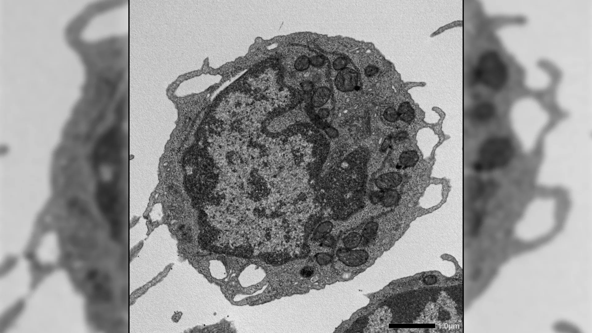
Researchers in Brazil are advancing a promising form of cancer immunotherapy by making natural killer (NK) cells more powerful and precise. In a recent study, scientists at the Ribeirão Preto Blood Center and the Center for Cell-Based Therapy (CTC) used the NK-92 cell line to test new designs of chimeric antigen receptors (CARs). These engineered receptors included specific costimulatory components, such as 2B4 and DAP12, that help activate the cells. The findings showed that these additions made the cells “ready to attack,” significantly improving their ability to destroy tumor cells. The study was published in Frontiers in Immunology.

CAR-based therapies have already transformed cancer treatment, particularly for blood-related cancers. While CAR-T cells are well studied, scientists are still working to understand how to optimize CAR-NK cells. One key challenge is identifying which internal signaling mechanisms allow these cells to perform at their best.

The new research addresses this gap by focusing on how specific signaling domains influence NK cell activity. By incorporating 2B4 and DAP12 into the CAR design, the researchers were able to enhance the cells’ activation state, making them more effective at targeting tumors.

Combining activation signals with drug control

The team also explored a strategy to fine-tune the cells using a temporary drug-based approach. They tested dasatinib, a drug that can briefly suppress cell activity, to see how controlled pauses might affect performance.

Their results suggest that combining optimized activation signals with reversible pharmacological control can improve both the strength and efficiency of CAR-NK therapies. This approach may help researchers design more advanced and controllable cell-based cancer treatments in the future.

Stronger tumor control in preclinical models

According to the Ribeirão Preto Blood Center Press Office, experiments in animal models showed encouraging results. CAR-NK cells engineered with 2B4-DAP12 and pretreated with dasatinib were better at controlling tumor growth compared to more traditional versions of the therapy.

Research collaboration and institutional support

The Center for Cell-Based Therapy (CTC) is one of the Research, Innovation, and Dissemination Centers (RIDCs) supported by FAPESP. It operates within the Ribeirão Preto Blood Center and is affiliated with the general and teaching hospital (“Hospital das Clínicas”) of the Ribeirão Preto Medical School of the University of São Paulo (FMRP-USP).

Together, these findings point toward a new generation of CAR-NK therapies that could offer stronger, more adaptable ways to fight cancer.

Share Button

A “death” protein may be the key to slowing aging at its source

As people get older, their blood and immune systems gradually lose strength. A major reason is the decline of hematopoietic stem cells (HSCs), which are responsible for producing all types of blood cells. Under healthy conditions, these stem cells can renew themselves and create a balanced mix of blood cells. Over time, however, they become less efficient. They generate fewer new cells, begin to favor certain types such as myeloid cells over lymphoid cells, and are less capable of supporting a strong immune response.

Several factors appear to drive this decline, including accumulated cellular damage, changes in gene activity, chronic low-level inflammation, and shifts in the bone marrow environment. Even so, scientists have not fully understood how these different stresses combine to impair HSC function.

Investigating a Key Aging Pathway

To better understand this process, researchers from The University of Tokyo, Japan, and St. Jude Children’s Research Hospital, USA, explored how age-related stress affects HSCs. They focused on the receptor-interacting protein kinase 3 (RIPK3)-mixed lineage kinase like (MLKL) signaling axis, which is typically associated with necroptosis, a form of programmed cell death.

The study was led by Dr. Masayuki Yamashita, an Assistant Member at St. Jude Children’s Research Hospital, who, at the time of the investigation, was an Assistant Professor at The Institute of Medical Science, The University of Tokyo. Co-authors included Dr. Atsushi Iwama from The Institute of Medical Science, The University of Tokyo, and Dr. Yuta Yamada from St. Jude Children’s Research Hospital, who was a graduate student at The Institute of Medical Science, The University of Tokyo.

A Surprising Discovery About MLKL

The research began with an unexpected observation. Dr. Yamashita explains, “We discovered an unexpected phenotype in HSCs of MLKL-knockout mice repeatedly treated with 5-fluorouracil, where aging-associated functional changes were markedly attenuated despite no detectable difference in HSC death, prompting us to investigate whether this pathway might induce functional changes beyond cell death.”

This finding suggested that MLKL might influence stem cell aging without actually killing the cells. That idea became central to the study, which was published in Volume 17 of Nature Communications on April 6, 2026.

How Scientists Tested the Mechanism

To explore this possibility, the researchers used several types of genetically engineered mice, including wild-type, MLKL-deficient, and RIPK3-deficient models. They also used specialized reporter mice designed to detect MLKL activation using a Förster resonance energy transfer-based biosensor.

The mice were exposed to different stress conditions that mimic aging, such as inflammation, replication stress, and oncogenic stress. To measure how well HSCs functioned, the team relied mainly on bone marrow transplantation, which tests the ability of stem cells to rebuild the blood system.

Additional techniques provided deeper insights, including flow cytometry, ex vivo expansion, RNA-seq, assay for transposase-accessible chromatin-seq, high-resolution imaging, metabolic testing, and detailed studies of mitochondria. Together, these approaches allowed the researchers to examine how MLKL affects HSCs at multiple levels.

Mitochondrial Damage Without Cell Death

The results revealed a previously unknown role for MLKL in stem cell aging. Although MLKL is usually linked to cell death, its activation in HSCs did not increase cell death or reduce cell numbers. Instead, it acted in a different way.

When activated under stress, MLKL briefly moved to the mitochondria, the structures that generate energy within cells. There, it caused damage by lowering membrane potential, altering mitochondrial structure, and reducing energy production. These effects led to key features of aging in HSCs, including reduced ability to renew themselves, decreased production of lymphoid cells, and a shift toward myeloid cell output.

Blocking MLKL Preserves Stem Cell Function

When MLKL was removed or inactivated, many of these problems were significantly reduced. HSCs lacking MLKL retained their ability to regenerate, produced healthier immune cells, showed less DNA damage, and maintained better mitochondrial function. These benefits were seen even in older animals or under stressful conditions.

Notably, these improvements occurred without major changes in gene expression or chromatin accessibility. This suggests that MLKL influences aging through processes that occur after gene activity, particularly at the level of cellular structures like mitochondria, rather than through changes in DNA regulation or inflammation.

Implications for Aging and Future Therapies

The findings point to a common pathway that connects various types of cellular stress to mitochondrial damage and stem cell aging. By identifying MLKL as a key link in this process, the study offers new insight into how aging affects the blood system.

Dr. Yamashita emphasizes, “In the longer term, this research could lead to therapies that preserve the function of hematopoietic stem cells, ultimately improving recovery and long-term health for patients undergoing chemotherapy, radiation, or transplantation. By revealing how non-lethal activation of cell-death pathways drives stem cell aging, these findings may inspire new classes of mitochondrial-protective or necroptosis-modulating drugs.”

A New Understanding of Stem Cell Aging

Overall, the study reveals that MLKL plays an important role in stem cell aging without causing cell death. Instead, it responds to stress by damaging mitochondria and weakening HSC function over time. This discovery challenges traditional views of necroptosis-related proteins and opens new possibilities for slowing or preventing age-related decline in the blood and immune systems.

Share Button

Lawmakers clash with RFK Jr as he shifts focus away from vaccines

During a three-hour hearing, the US health secretary tried to focus on chronic disease while being pressed on vaccines.

Share Button

Rollout of Covid vaccines extraordinary feat – inquiry report

Covid vaccines saved hundreds of thousands of lives, but a small minority harmed need better support, says report.

Share Button

Vaccines a huge success, but public trust must be earned – key findings from Covid report

Immunisation saved hundreds of thousands of UK lives, but vaccine hesitancy remains an issue.

Share Button

Breakthrough £90,000 Alzheimer’s drugs unlikely to benefit patients, report suggests

A major review has provoked a backlash after concluding the medicines provide too little benefit to be noticed.

Share Button

I feared my son had a brain tumour but he’d been poisoned with vitamin D

Investigations found Roo had been accidentally poisoned with a dose of vitamin D prescribed for growing pains.

Share Button

Wellbeing garden opens to combat isolation

The outdoor area at Yarm Wellness will officially open to the public on Saturday.

Share Button

Senior midwife appointed to maternity inquiry

Senior midwife Donna Ockenden will review maternity services in Sussex after a campaign by families.

Share Button

A crushed fossil revealed a dinosaur that shouldn’t have existed

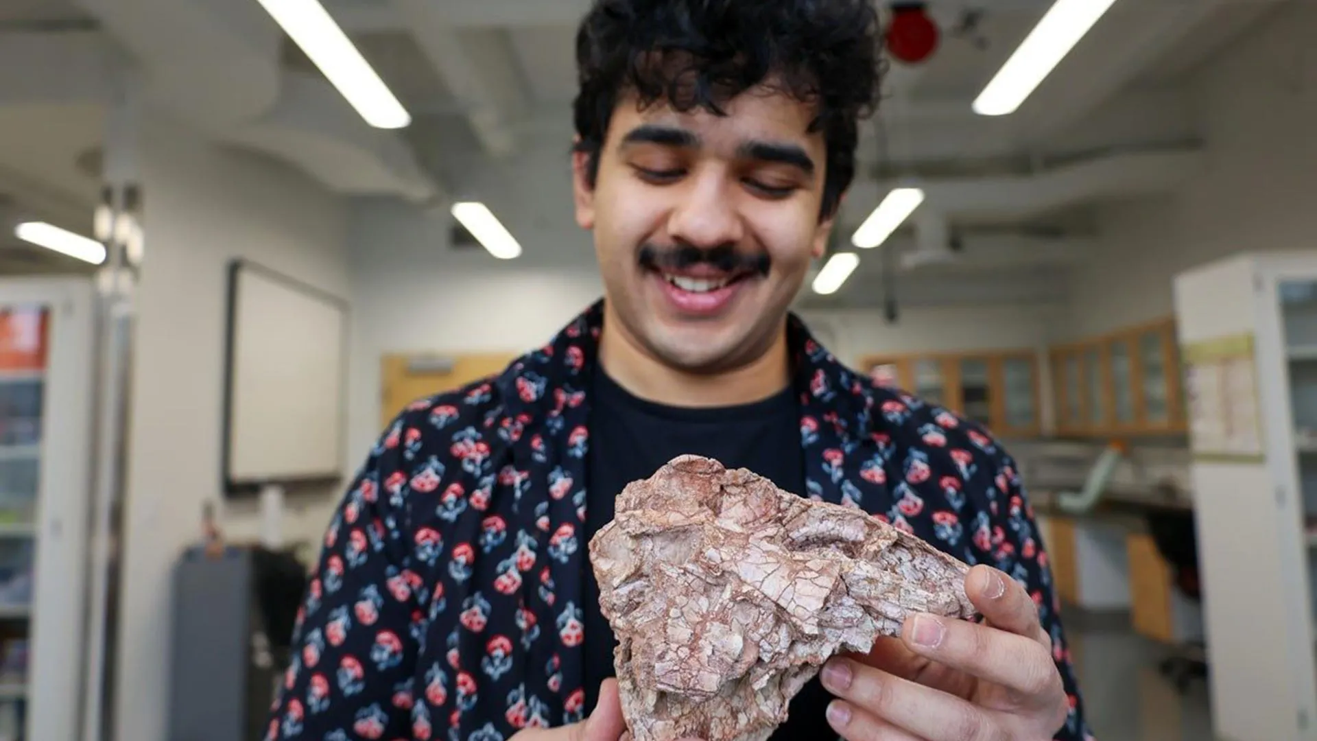
“You want to stick your finger in a dinosaur brain?” asked Simba Srivastava.

Inside a paleobiology lab lined with cabinets of ancient fossils, the Virginia Tech undergraduate held up a rough, pitted skull.

“This is a uniquely sucky specimen,” said Srivastava. “It’s so bad. Like, if you saw a human skull in this way, you’d throw up.”

Despite its poor condition, the senior geosciences major spent two years carefully reconstructing the fossil and figuring out where it fits in the evolutionary history of dinosaurs. His work, published in Papers in Palaeontology, offers new insight into how dinosaurs rose to dominance during the Jurassic period.

Although this kind of research is typically handled by experienced scientists, geobiologists Sterling Nesbitt and Michelle Stocker brought Srivastava onto the project as a first-year student.

“We want undergraduate researchers to experience the whole paleontological research process at Virginia Tech,” said Nesbitt. “Simba grabbed the project by the reins.”

Reconstructing a Rare Dinosaur Skull

The fossil had an unusual history. It was first discovered in 1982 by a team from the Carnegie Museum of Natural History at Ghost Ranch in New Mexico. More than 30 years later, Nesbitt rediscovered it in a drawer and brought it to Virginia Tech for further study.

Using computed tomography scanning data, Srivastava digitally separated the crushed bones and created a 3D printed reconstruction of the skull.

The fossil belonged to a carnivorous dinosaur species that lived more than three times earlier than Tyrannosaurus Rex.

These animals lived near the end of the Triassic period, which lasted from about 252 million to 201 million years ago. At that time, dinosaurs were not yet the dominant predators often seen in movies. They competed with early relatives of crocodiles and mammals for survival.

How Dinosaurs Rose to Power

That balance changed dramatically after a mass extinction event eliminated much of the competition. As the Triassic period ended, dinosaurs quickly became the dominant land animals.

“Dinosaurs go from being co-stars to the headliner,” Srivastava said.

Fossils from this critical transition are rare, especially well-preserved ones from the end of the Triassic. That makes this damaged skull especially valuable.

In fact, no other specimen like it has been found.

Even in its distorted state, the fossil revealed important details. The dinosaur had large cheekbones, a broad braincase, and likely a short, deep snout. These features had not been seen before in early dinosaurs, suggesting they were evolving in more complex ways than previously understood.

A New Species With a Strange Look

Srivastava named the new species based on its unusual appearance.

“We landed on Ptychotherates bucculentus, which means ‘folded hunter with full cheeks’ in Latin,” said Srivastava. “One paleo-artist said that it looked like a murder muppet.”

After years of analysis, the team determined that this dinosaur belonged to Herrerasauria, one of the earliest groups of carnivorous dinosaurs. It appears to have been among the last surviving members of this lineage.

Rethinking the End-Triassic Extinction

The fossil led to another unexpected conclusion.

Ptychotherates was found in rock layers that may date to just before the mass extinction at the end of the Triassic period, and no other members of its group have been found after that time, possibly suggesting that this dinosaur group went extinct as a result of that mass extinction.

“This forces us to reconsider the impact of the end-Triassic extinction as something that wiped out not just the competitors to dinosaurs, but some long-standing dinosaur lineages themselves,” Srivastava said.

Because no herrerasaurians have been discovered elsewhere from such a late point in the Triassic, scientists think the region that is now the American Southwest may have been their final refuge.

One Fossil, an Entire Lost Lineage

Srivastava’s “folded hunter” may be the only remaining evidence of this group’s final chapter.

“This specimen, it fits in my hands, but it is the only proof that any of these dinosaurs lived this long, lived in these latitudes, the only proof that they evolved to have this skull shape,” said Srivastava. “All these billions of individuals that existed through time are spoken for by this one specimen.”

Share Button