Scientists discover hidden driver of aging — Simple supplement reversed brain decline

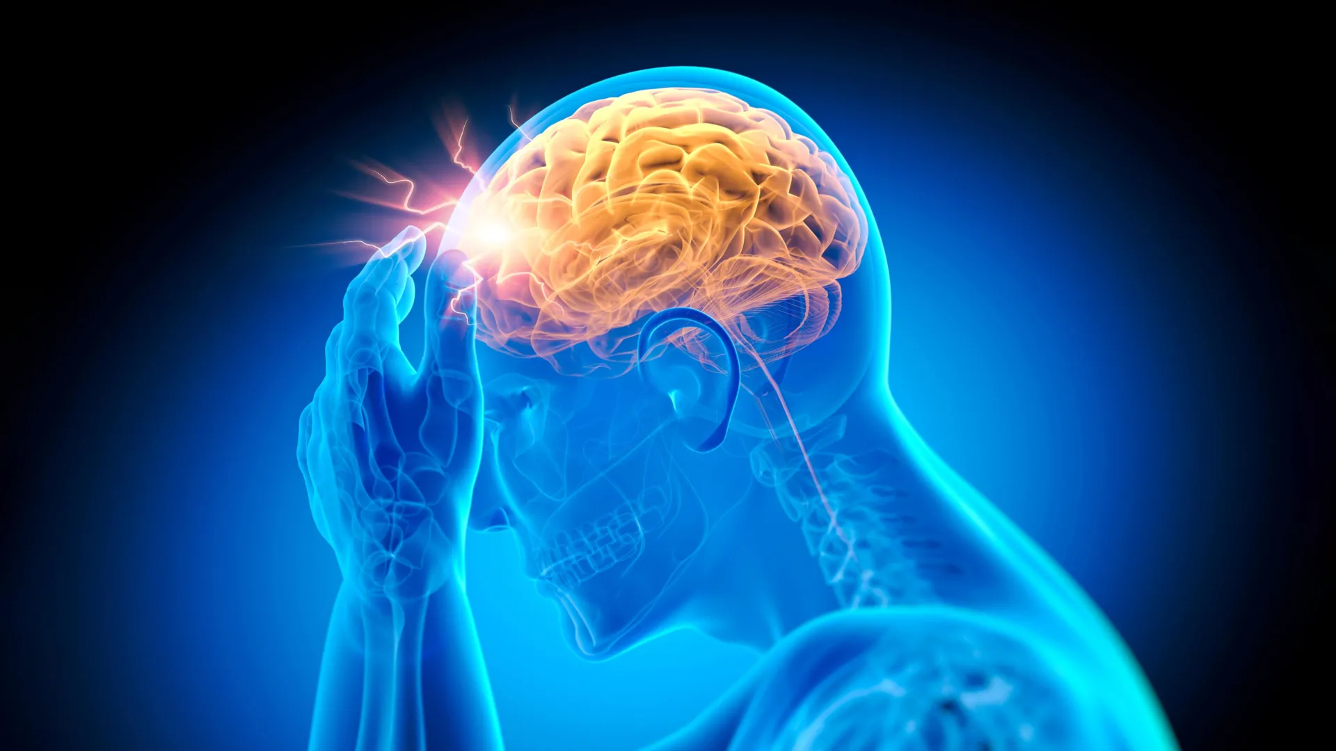
Scientists may have uncovered a hidden biological switch that helps control how quickly the body ages. Research published in PLOS Biology suggests that declining levels of a brain protein called Menin can trigger inflammation, memory decline, and other age-related changes throughout the body. In experiments with mice, restoring the protein reversed several signs of aging, while a simple amino acid supplement improved cognitive function.

The findings add to growing evidence that aging may be strongly influenced by the hypothalamus, a small but powerful brain region that regulates metabolism, hormones, body temperature, sleep, and stress responses. Researchers increasingly view the hypothalamus as a central command center for aging itself.

A Brain Protein That Declines With Age

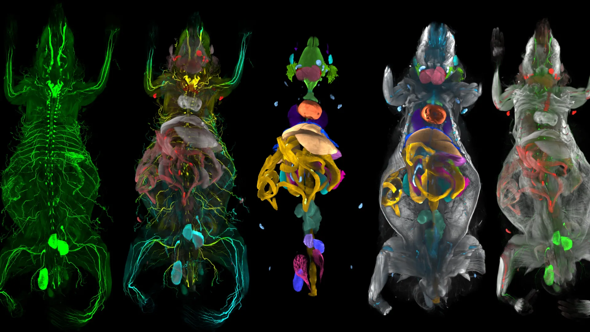
The study, led by Lige Leng and colleagues at Xiamen University in China, focused on Menin, a protein that helps suppress inflammation in the brain. Earlier work had already shown that Menin plays an important role in controlling neuroinflammatory activity. The team wanted to know whether losing this protective protein might contribute to aging.

Their experiments revealed that Menin levels dropped sharply in the hypothalamus as mice grew older. The decline occurred specifically in neurons within the ventromedial hypothalamus (VMH), a region linked to metabolism and systemic aging. Interestingly, Menin levels did not significantly decrease in nearby support cells such as astrocytes or microglia.

To investigate what this loss might mean, the researchers engineered mice in which Menin activity could be selectively reduced. The effects were striking. Younger mice with lower Menin levels developed increased brain inflammation, thinning skin, lower bone mass, impaired balance, memory problems, and a shorter lifespan compared with normal mice.

The results suggest that Menin may act as a protective “anti-aging” factor inside the brain.

The D-Serine Connection

One of the most surprising discoveries involved D-serine, an amino acid that also functions as a neurotransmitter in the brain. D-serine helps regulate communication between neurons and is important for learning and memory.

When Menin levels fell, D-serine production also dropped. The researchers traced this effect to reduced activity of an enzyme required for D-serine synthesis, which itself appears to be regulated by Menin.

D-serine naturally occurs in foods including soybeans, eggs, fish, and nuts, and it is also sold as a dietary supplement.

The connection caught researchers’ attention because other studies have linked declining D-serine levels with aging-related cognitive impairment and reduced synaptic plasticity, the brain’s ability to strengthen neural connections involved in memory and learning.

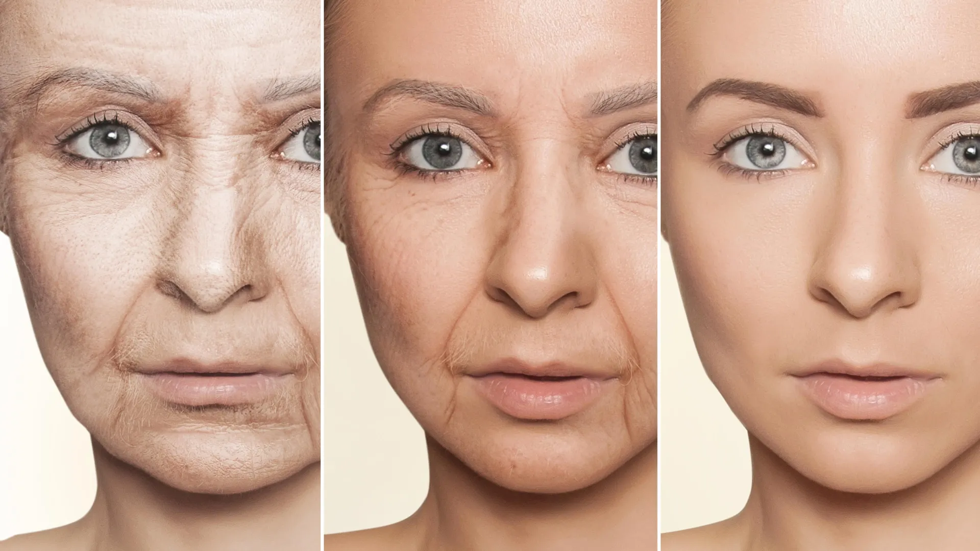
Reversing Signs of Aging in Mice

The researchers then tested whether restoring Menin could reverse age-related decline.

They delivered the Menin gene directly into the hypothalamus of elderly mice that were about 20 months old, roughly equivalent to late-life aging in humans. Just 30 days later, the animals showed measurable improvements in learning, memory, balance, skin thickness, and bone density.

The improvements were accompanied by increased D-serine levels in the hippocampus, a brain region essential for memory formation.

The team also tested whether D-serine supplementation alone could help. After three weeks of supplementation, older mice displayed better cognitive performance, although the treatment did not reverse the physical aging markers seen in skin and bone tissue.

That distinction suggests Menin likely affects aging through several interconnected biological pathways, not just D-serine production alone.

Why the Hypothalamus Is Becoming a Major Focus in Aging Research

Interest in the hypothalamus has grown rapidly in recent years as scientists uncover evidence that this brain region may coordinate many aspects of aging throughout the body.

More recent research has explored how age-related changes in hypothalamic DNA methylation and hormone signaling could contribute to neurodegenerative diseases such as Alzheimer’s. One 2024 study in Nature Communications found that the hypothalamus undergoes distinctive epigenetic changes with age and may influence pathways involving oxytocin and gonadotropin-releasing hormone (GnRH), both linked to aging and brain health.

Together, these findings strengthen the idea that aging is not simply the result of wear and tear across the body. Instead, some scientists suspect the brain may actively regulate parts of the aging process through inflammation, metabolism, and hormonal signaling.

Could D-Serine Help Humans?

Despite the excitement surrounding the findings, the research remains early and was conducted in mice, not humans. Scientists still do not know whether boosting Menin or supplementing with D-serine could safely slow aging or improve cognition in people.

Researchers also caution that altering powerful brain signaling pathways could have unintended consequences. More work is needed to understand why Menin declines with age, how long any benefits might last, and whether D-serine supplementation could produce side effects over time.

Still, the study offers an intriguing glimpse into how aging may one day be targeted more directly.

Leng said, “We speculate that the decline of Menin expression in the hypothalamus with age may be one of the driving factors of aging, and Menin may be the key protein connecting the genetic, inflammatory, and metabolic factors of aging. D-serine is a potentially promising therapeutic for cognitive decline.”

Leng also noted, “Ventromedial hypothalamus (VMH) Menin signaling diminished in aged mice, which contributes to systemic aging phenotypes and cognitive deficits. The effects of Menin on aging are mediated by neuroinflammatory changes and metabolic pathway signaling, accompanied by serine deficiency in VMH, while restoration of Menin in VMH reversed aging-related phenotypes.”

Share Button

Scientists warn popular vitamin D supplement may have a “previously unknown” downside

Taking vitamin D2 might lower the body’s levels of the more efficient form of vitamin D, vitamin D3, according to new research from the University of Surrey, John Innes Centre and Quadram Institute Bioscience. Many people take vitamin D supplements to support their bone and immune health and meet the UK government recommendation of 10 micrograms (µg) each day, especially during the winter months. 

There are two forms of vitamin D supplements available: vitamin D2 and vitamin D3. Researchers have found that taking vitamin D2 supplements can lead to a drop in the body’s concentration of vitamin D3, which is the form our bodies naturally produce from sunlight and use most effectively to raise overall vitamin D levels.  

The study, published in Nutrition Reviews, analyzed data from randomized controlled trials and found that vitamin D2 supplementation resulted in a reduction in vitamin D3 levels compared to those not taking a vitamin D2 supplement. In many of the studies, the vitamin D3 levels went lower than in the control group. 

Emily Brown, PhD Research Fellow and Lead Researcher of the study from the University of Surrey’s Nutrition, Exercise, Chronobiology & Sleep Discipline, said: 

“Vitamin D supplements are important, especially between October and March, when our bodies cannot make vitamin D from sunlight in the UK.  However, we discovered that vitamin D2 supplements can actually decrease levels of vitamin D3 in the body, which is a previously unknown effect of taking these supplements. This study suggests that subject to personal considerations, vitamin D3 supplements may be more beneficial for most individuals over vitamin D2.”  

Professor Cathie Martin, Group Leader at the John Innes Centre, said:  

“This meta-analysis highlights the importance of ensuring plant-based vitamin D3 is accessible in the UK.” 

This research supports a previous study published in Frontiers in Immunology, led by Professor Colin Smith from the University of Surrey, which suggests that vitamin D2 and D3 do not have identical roles in supporting immune function. Vitamin D3 has a modifying effect on the immune system that could fortify the body against viral and bacterial diseases.   

Professor Colin Smith said: 

“We have shown that vitamin D3, but not vitamin D2, appears to stimulate the type I interferon signalling system in the body – a key part of the immune system that provides a first line of defence against bacteria and viruses. Thus, a healthy vitamin D3 status may help prevent viruses and bacteria from gaining a foothold in the body.” 

Further research into the different functionalities of vitamin D2 and D3 should be a priority in deciding whether vitamin D3 should be the first-line choice of vitamin D supplement, subject to individual requirements. 

Professor Martin Warren, Chief Scientific Officer at the Quadram Institute, said: 

 “Vitamin D deficiency represents a significant public health concern, especially during the winter months with significant deficiency across the UK population. This collaborative research effort aligns well with the Quadram Institute’s mission to deliver healthier lives through food innovation to enhance the nutrient density of the food we eat. Tackling this with the most effective form of vitamin D supplementation or fortification is of the utmost importance to the health of the nation.” 

Share Button

New AI body map reveals obesity’s hidden attack on facial nerves

Researchers at Helmholtz Munich, Ludwig Maximilians University Munich (LMU), and several partner institutions have created an artificial intelligence (AI) system capable of mapping disease-related changes throughout an entire mouse body at cellular-level detail. Using the new platform, known as MouseMapper, the team discovered widespread inflammation and previously unknown nerve damage linked to obesity.

The study also identified similar molecular patterns in human tissue, suggesting that important aspects of obesity-related nerve damage may occur in both mice and people. The findings were published in the journal Nature.

Obesity is known to affect much more than body weight and metabolism. It can alter immune activity, disrupt nerve structures, and reshape tissues throughout the body, increasing the risk of conditions such as type 2 diabetes, cardiovascular disease, stroke, neuropathy, and cancer. Despite these widespread effects, scientists have lacked tools capable of studying disease-related changes across an entire intact body in high detail.

To address that challenge, a research team led by Prof. Ali Ertürk, Director of the Institute for Biological Intelligence (iBIO) at Helmholtz Munich and Professor at LMU, developed MouseMapper. The AI framework uses foundation-model-based deep learning algorithms to analyze massive whole-body imaging datasets.

The system can automatically identify and segment 31 organs and tissue types while also mapping nerves and immune cells throughout the body. This allows researchers to examine how diseases affect multiple organ systems at the same time in intact mice.

“MouseMapper is built on a foundation model, which means it generalizes far beyond the data it was originally trained on,” says Ying Chen, co-first author of the study.

Transparent Mice and Whole-Body Imaging

To build the body maps, researchers first tagged nerves and immune cells in mice using fluorescent markers that glow under a microscope. They then used tissue-clearing methods to make the mice transparent while preserving the fluorescent signals, allowing scientists to see deep inside the body without cutting tissues apart.

Next, the team used advanced light-sheet microscopy to capture detailed three-dimensional images of entire mice. The process generated enormous datasets containing tens of millions of cellular structures from organs and tissues across the body.

MouseMapper then analyzed the images automatically, identifying anatomical regions, nerve networks, and immune-cell clusters throughout the animals.

This approach allowed the researchers to pinpoint exactly where inflammation and tissue damage appeared in organs such as fat tissue, muscle, liver, and peripheral nerves. Unlike earlier methods, scientists did not need to choose specific regions to study beforehand.

Obesity Linked to Facial Nerve Damage

To explore how obesity changes the body, the researchers fed mice a high-fat diet that produced obesity and metabolic problems similar to those seen in humans.

Using MouseMapper, the team found widespread alterations in immune-cell organization and nerve structures across the body. One of the most surprising discoveries involved the trigeminal nerve, a major facial nerve responsible for facial sensation and certain motor functions.

In obese mice, these sensory nerves showed a major reduction in branches and nerve endings, suggesting impaired nerve function. Behavioral tests supported that conclusion, showing that obese mice were less responsive to sensory stimulation compared to lean mice.

The researchers then focused on the trigeminal ganglion, which contains the cell bodies of facial sensory neurons. Through spatial proteomics analysis, they identified molecular changes linked to inflammation and nerve remodeling.

Importantly, many of the same molecular signatures were also found in trigeminal tissue from people with obesity. This suggests that the nerve-related changes observed in mice may also occur in humans.

“We revealed previously unknown structural and molecular changes in the trigeminal ganglion and its facial branches, and the same molecular signature was conserved in human tissue. This kind of finding simply cannot emerge from studying one organ at a time,” says Dr. Doris Kaltenecker, senior scientist at the Institute for Diabetes and Cancer (IDC) at Helmholtz Munich and first author of the study.

A New Tool for Studying Complex Diseases

The researchers believe MouseMapper could become an important tool for studying diseases that affect many organ systems simultaneously, including diabetes, cancer, neurodegenerative diseases, and autoimmune disorders.

Unlike earlier approaches focused on individual tissues or organs, MouseMapper provides an integrated whole-body analysis system that can identify disease hotspots throughout an organism.

The team has also made the whole-body datasets publicly available online so researchers around the world can explore obesity-related changes across organs and tissues.

“Our goal is to create a comprehensive framework for understanding how diseases affect the body as an interconnected system,” says Ali Ertürk. “Our long-term vision is to build truly realistic digital twins of mice in health and disease: cell-level atlases that we can query, perturb and screen in silico computationally. That would let us pinpoint the earliest changes a disease causes, design interventions to prevent them, and accelerate the discovery of new treatments while reducing the number of physical experiments we need to run.”

The work was supported by the European Research Council (Consolidator Grant CALVARIA to A. Ertürk; grant 949017 to M. Rohm), the German Research Foundation (DFG) under Germany’s Excellence Strategy within the Munich Cluster for Systems Neurology (SyNergy, ID 390857198, EXC 2145), DFG SFB 1052 (A9) and TR 296 (P03), the Collaborative Research Centre CRC 1744, the German Federal Ministry of Education and Research (NATON collaboration, 01KX2121, and HIVacToGC), the Vascular Dementia Research Foundation, the Nomis Heart Atlas Project Grant (Nomis Foundation), the Else-Kröner-Fresenius-Stiftung, the Edith-Haberland-Wagner Stiftung, the Helmut Horten Foundation, the EFSD and Novo Nordisk A/S Programme for Diabetes Research in Europe (to D. Kaltenecker), and the China Scholarship Council (to Y. Chen).

Share Button

Excessive social media ‘negatively impacts wellbeing’

The World Happiness Reports finds the more time spent on social media the greater loss of wellbeing.

Share Button

AI may speed up search for drugs to treat brain conditions

Researchers hope the work will help identify affordable, effective drugs to treat conditions like MND.

Share Button

Ebola risk raised to ‘very high’ in DR Congo

The head of the UN health agency says the risk in the wider region is “high”, but it remains “low” at the global level.

Share Button

‘People can feel lonely in a crowd’

Dawn French discusses loneliness, particularly among older women, on Woman’s Hour.

Share Button

UK scientists developing Ebola vaccine that could be ready for trials in months

The rare species of Ebola involved – known as Bundibugyo – kills around a third of those infected and has no proven vaccine yet.

Share Button

Scientists uncover cancer-causing chemicals hidden in everyday foods

More people are paying close attention to what they eat, often tracking calories, exercising daily, and filling their plates with foods that seem naturally healthy, including fruits and vegetables. Yet even nutritious foods can carry hidden chemical concerns. Some contaminants can enter food from the environment, while others can form during high heat cooking methods such as heating, smoking, grilling, roasting, and frying.

Among the compounds of concern are polycyclic aromatic hydrocarbons, or PAHs (hydrophobic organic compounds comprising multiple fused aromatic rings). Some PAHs are known for their cancer causing potential, which makes reliable food testing an important part of protecting public health.

A Hidden Food Safety Challenge

Detecting PAHs in food is not simple. Conventional extraction methods, such as solid phase extraction, liquid liquid extraction, and accelerated solvent extraction, can be affordable, but they often require lengthy preparation, heavy hands on labor, and chemical intensive procedures that are not ideal for workers or the environment.

To solve these problems, scientists have been turning to a streamlined method known as QuEChERS (Quick, Easy, Cheap, Effective, Rugged, and Safe). The approach is designed to speed up sample preparation, reduce chemical use, improve recovery rates, and make food contaminant testing more practical for routine safety checks.

In a 2025 study, researchers from the Department of Food Science and Biotechnology at Seoul National University of Science and Technology, led by Professor Joon-Goo Lee, used QuEChERS to measure eight PAHs (Benzo[a]anthracene, Chrysene, Benzo[b]fluoranthene, Benzo[k]fluoranthene, Benzo[a]pyrene, Indeno[1,2,3-cd]pyrene, Dibenz[a,h]anthracene, and Benzo[g,h,i]perylene in food. The findings were published in the journal Food Science and Biotechnology.

Faster Testing With Strong Accuracy

The team used acetonitrile to extract PAHs from food samples, then tested several purification strategies involving different combinations of sorbents. The method was validated across multiple food matrices, showing strong performance. Calibration curves for all eight PAHs had R2 values above 0.99, indicating a highly linear and reliable measurement system.

Further analysis using gas chromatography and mass spectrometry showed that the limits of detection ranged from 0.006 to 0.035 µg/kg, while the limits of quantification ranged from 0.019 to 0.133 µg/kg. Recovery rates were also strong, ranging from 86.3 to 109.6% at 5 µg/kg, 87.7 to 100.1% at 10 µg/kg, and 89.6 to 102.9% at 20 µg/kg. Precision values stayed between 0.4 and 6.9% across all tested food matrices.

The study also reported that, among the foods tested, the highest PAH levels were found in soybean oil, followed by duck meat and canola oil.

Prof. Lee explains, “This method not only simplifies the analytical process but also demonstrates high efficiency in detection compared to conventional methods. It can be applied to a wide range of food matrices.”

Why PAHs Matter

PAHs can form when food is exposed to high temperatures or smoke. According to the National Cancer Institute, PAHs can develop when fat and juices from meat drip onto a hot surface or open flame, creating smoke that deposits these compounds onto the food. PAHs can also form during smoking and may be found in sources such as cigarette smoke and car exhaust fumes.

The NCI notes that PAHs and related high temperature cooking compounds have caused cancer in animal studies, although human population studies have not established a definitive link between exposure from cooked meats and cancer. This uncertainty is one reason more accurate measurement tools are valuable. Better testing can help regulators, researchers, and food companies understand where contamination is occurring and how it can be reduced.

Newer Research Points to Broader Use

Since the SeoulTech study, other researchers have continued refining QuEChERS based methods for PAH detection. A 2025 study in Foods developed a modified QuEChERS method with a freeze out step and applied it to 302 retail food samples. That work found the highest concentration of four priority PAHs in Kezuribushi, a smoked and dried fish product, and identified grilled chicken feet as a possible health concern based on the European Food Safety Authority margin of exposure approach.

Another 2025 study focused on cereals and cereal based products. Researchers developed a modified QuEChERS method using Z Sep⁺ clean up and gas chromatography with tandem mass spectrometry. In 96 cereal samples and 18 cereal based products from the Romanian market, only chrysene was quantified in 17% of cereal samples, while no PAHs were quantified in the derived products.

Together, these newer findings suggest that QuEChERS based approaches are becoming increasingly useful for different food categories, from oils and meats to smoked products and cereals. They also show why food specific testing matters, since PAH levels can vary widely depending on ingredients, processing, cooking methods, and environmental exposure.

Safer Food Testing and Cleaner Labs

For the food industry, a faster and more efficient PAH testing method could improve safety management by making it easier to inspect products before they reach consumers. The approach may also reduce costs and improve working conditions by cutting down on time consuming procedures and limiting the use of hazardous chemicals.

“Our research can improve public health by providing safe food. It also reduces the use and emission of hazardous chemicals in laboratory testing,” concludes Prof. Lee.

The broader takeaway is clear: food safety testing is becoming faster, cleaner, and more precise. By improving how scientists detect PAHs, methods like QuEChERS could help identify hidden contaminants, support safer food production, and reduce chemical waste in the lab.

About Professor Joon Goo Lee

Joon Goo Lee is a Professor at the Department of Food Science and Biotechnology, Seoul National University of Science and Technology. He is an expert in food regulation and safety assessment. He served as a scientific officer at Korea’s Ministry of Food and Drug Safety and as a visiting researcher at FSANZ. He is a member of the National Food Sanitation Committee and an expert for the FAO/WHO JECFA. He also serves as the executive director of the Korean food safety societies. His research focuses on risk assessment and the reduction of contaminants in food, contributing to science based policies and improved public health.

Share Button

Scientists warn that current vitamin B12 guidelines may be putting your brain at risk

Vitamin B12 is best known for helping the body make DNA, red blood cells, and healthy nerve tissue. But research suggests that simply meeting the current minimum standard may not always be enough, especially for older adults.

A UCSF led study found that healthy older people with lower vitamin B12 levels, even when those levels still fell within the accepted normal range, showed signs of subtle neurological and cognitive problems. The findings raise a provocative possibility: some people may be told their B12 status is fine while their brains are already showing early signs of strain.

“Normal” B12 May Not Always Mean Optimal

The study, published in Annals of Neurology, looked at older adults who did not have dementia or mild cognitive impairment. Even in this relatively healthy group, lower levels of active B12 were linked to slower thinking, slower visual processing, and more visible injury in the brain’s white matter. White matter is made up of the nerve fibers that allow different parts of the brain to communicate.

The work was led by senior author Ari J. Green, MD, of the UCSF Departments of Neurology and Ophthalmology and the Weill Institute for Neurosciences. Green and his colleagues said the results call attention to a possible weakness in current B12 guidelines. The minimum threshold used to define deficiency may not capture early functional changes in the nervous system.

“Previous studies that defined healthy amounts of B12 may have missed subtle functional manifestations of high or low levels that can affect people without causing overt symptoms,” said Green, noting that clear deficiencies of the vitamin are commonly associated with a type of anemia. “Revisiting the definition of B12 deficiency to incorporate functional biomarkers could lead to earlier intervention and prevention of cognitive decline.”

Brain Scans Revealed a Troubling Pattern

Researchers enrolled 231 healthy participants through the Brain Aging Network for Cognitive Health (BrANCH) study at UCSF. The participants had an average age of 71, and none had dementia or mild cognitive impairment.

Their average blood B12 level was 414.8 pmol/L, far above the U.S. minimum cutoff of 148 pmol/L. Instead of relying only on total B12, the researchers focused on the biologically active form of the vitamin, which may better reflect how much B12 the body can actually use.

After adjusting for age, sex, education, and cardiovascular risk factors, the team found that participants with lower active B12 had slower processing speed on cognitive tests. The effect was stronger with older age. They also had delayed responses to visual stimuli, pointing to slower visual processing and reduced brain signaling efficiency.

MRI scans added another warning sign. Participants with lower active B12 had a higher volume of white matter lesions, which are areas of brain injury that have been linked to cognitive decline, dementia, and stroke risk.

Why Older Adults May Be More Vulnerable

The study focused on older adults, a group that may be especially sensitive to lower B12 because absorption can become less efficient with age. Some medications, digestive conditions, and diets low in animal based foods can also increase the risk of low B12.

Co-first author Alexandra Beaudry-Richard, MSc, said the findings suggest that low but technically normal B12 could have broader effects than previously recognized. These levels could “impact cognition to a greater extent than what we previously thought, and may affect a much larger proportion of the population than we realize.” Beaudry-Richard is currently completing her doctorate in research and medicine at the UCSF Department of Neurology and the Department of Microbiology and Immunology at the University of Ottawa.

“In addition to redefining B12 deficiency, clinicians should consider supplementation in older patients with neurological symptoms even if their levels are within normal limits,” she said. “Ultimately, we need to invest in more research about the underlying biology of B12 insufficiency, since it may be a preventable cause of cognitive decline.”

Newer Evidence Adds Important Context

Research published after and around the UCSF study has added nuance rather than a simple answer. A 2025 comprehensive review concluded that B12 deficiency remains a modifiable risk factor for neurological and cognitive problems, especially in high risk groups such as older adults and vegetarians. The review also highlighted the growing importance of better biomarkers and brain imaging for detecting problems earlier.

A 2025 systematic review and meta analysis of randomized trials found that supplementation with B vitamins, including B6, B9, or B12, produced a very small benefit in global cognitive function among older adults. The authors rated the cleaned analysis as high certainty, but the effect was small, suggesting that supplementation is not a dramatic brain boost for everyone.

Another 2025 study using Mendelian randomization found no clear evidence that genetically higher total serum B12 levels protect the general population from psychiatric disorders or cognitive impairment. However, the authors noted an important limitation: their analysis used total serum B12, not the bioactive form measured in the UCSF work.

Together, the newer evidence supports a more careful message. B12 is clearly essential for the nervous system, and deficiency should not be ignored. But simply raising B12 for everyone may not be the answer. The more urgent question is whether current testing misses people whose brains are already affected despite “normal” results.

A Preventable Risk Worth Taking Seriously

The UCSF findings do not prove that lower active B12 directly causes cognitive decline, and they do not mean every older adult should begin taking supplements without medical guidance. They do, however, suggest that the current definition of B12 deficiency may be too blunt for brain health.

For clinicians, the study points to the possible value of looking beyond total B12, especially when older patients have neurological symptoms. For patients, it highlights a practical message: a “normal” lab result may not always tell the full story, particularly when subtle changes in memory, thinking speed, or vision are already appearing.

Authors: Co first author is Ahmed Abdelhak, MD, PhD, of the UCSF Department of Neurology and the Weill Institute for Neurosciences.

Funding and Disclosures: Westridge Foundation and the Canadian Institutes of Health and Research. There are no conflicts of interest to report.

Share Button