Once-discounted binding mechanism may be key to targeting viruses

“Position 4” didn’t seem important until researchers took a long look at a particular peptide.

That part of the peptide drawn from a SARS-CoV virus turned out to have an unexpected but significant influence on how it stably binds with a receptor central to the immune system’s ability to attack diseased cells.

In a study published by the Proceedings of the National Academy of Sciences, researchers at Rice University’s Brown School of Engineering and the University of Texas MD Anderson Cancer Center revealed models at atomic resolution that detail not only the binding but also, for the first time, the unbinding mechanisms that underlie a key component of the immune system.

They say better understanding of the entire mechanism could lead to advancements in immunotherapy that boosts the body’s ability to fight disease.

Rice computer scientist Lydia Kavraki, alumnus Jayvee Abella and postdoctoral researcher Dinler Antunes, led the study.

“Finding good targets to trigger a protective immune response is very challenging, especially in cancer research,” Antunes said. “The fact that this particular peptide was predicted not to bind to HLAs (human leukocyte antigens) by sequence-based methods highlights a blind spot in our current prediction capacity.

“By incorporating structural analysis, we can detect the contribution of these secondary interactions to peptide binding and stability, hopefully enabling us to find better targets for antiviral vaccine development and T-cell-based cancer immunotherapy,” he said.

The researchers used their simulations to illuminate details of how the intracellular SARS peptide, QFKDNVILL, binds to an MHC receptor protein known as HLA-A?24:02, primarily at dominant anchors on both ends of the peptide (at positions 2 and 9) and presents them for inspection to the immune system’s T cells.

Stable binding of a peptide and MHC is a prerequisite to the activation of T cells, which look for peptides not normally found in healthy cells. If the peptide and protein don’t bind, the T cell is not prompted to attack.

“That much was known from previous studies of the bound and unbound states of many such complexes,” Kavraki said. “What they didn’t capture was the intermediate states and the transitions that lead from one state to another, especially the unbinding.

“I think this is the only analysis that shows the unbinding of peptides from the MHC with atomic resolution,” Kavraki said. “Other peptides have similar characteristics and we think they would have similar behaviors.”

All of these interactions were revealed in great detail through Markov state models that analyze how systems change over time. In this case, the models revealed the importance of secondary sites that support the peptide’s primary anchors. That’s where position 4 stood out.

“There are the main, canonical anchors that people know, but there are these secondary interactions that contribute to the binding and the stability,” Antunes said. “These are harder to capture, but in this study, it seems that position 4 plays a very important role. When you mutate it, it affects the behavior of the peptide as it unbinds from the molecule.”

The researchers modeled mutations of the MHC to see how they would influence binding and found they supported the importance of position 4 to the stability of the complex.

“Our computational approach was able to make predictions on the effect of mutations that are then experimentally verified,” said co-author Cecilia Clementi, a former Rice professor who recently became Einstein Professor of Physics at the Free University of Berlin.

The researchers developed a two-stage process to simplify the computational complexity of atom-scale analysis of large molecules. The first stage used a technique called umbrella sampling to accelerate the initial exploration of the molecules. The second, exploratory stage used adaptive sampling, in which simulations are driven to accelerate the construction of the Markov model.

“The challenge is that these MHCs are pretty large systems for computational chemists to simulate,” said Abella, whose research on the topic formed much of his doctoral thesis. “We had to make some approximations and leverage advances in these classes of methods to move forward.

“We’re not the first one to study unbinding, but what characterizes our work over others is that we keep full atomic resolution in our simulations,” he said. “Other works use a technique known as a Markov chain Monte Carlo, whereas we use molecular dynamics, which lets us incorporate time into our computation to capture the kinetics.”

Their methods can be applied to other peptide-MHC complexes with existing 3D models. “This was, in some sense, a feasibility study to show we can use molecular dynamics and build a Markov state model of a system this size,” Abella said.

The researchers also noted the study’s relevance to the current fight against COVID-19, as the SARS peptide they viewed, QFKDNVILL, is highly similar to the NFKDQVILL peptide in SARS-CoV-2, with the same binding pockets in positions 2, 4 and 9.

“These results suggest that both peptides can bind to HLA-A*2402 and provide targets for anti-viral T-cell responses, which are of great interest in light of the current pandemic,” said co-author Gregory Lizée, a professor in the Department of Melanoma Medical Oncology at MD Anderson. “But these results also shed light on many other potential immune targets, including those of other viruses and even human cancers.”

Kavraki noted that experimental work by longterm collaborator Lizée and Kyle Jackson, a graduate research assistant at Lizée’s lab who produced the mutant proteins, were critical to validate their simulations. Kavraki’s own lab won a National Science Foundation (NSF) Rapid Response Research grant to help identify fragments of SARS-CoV-2 viral proteins as possible targets for vaccine development.

Kavraki is the Noah Harding Professor of Computer Science and a professor of bioengineering, mechanical engineering and electrical and computer engineering.

The Cancer Prevention and Research Institute of Texas, the Gulf Coast Consortia, the NSF, the Einstein Foundation Berlin and the Welch Foundation supported the research.


Share Button

Novel insights on cellular suicide could provide new avenues for cancer therapies

When it comes to complex life — that of the multicellular variety — cell death can be just as important as survival. It allows organisms to clean house and prevent the proliferation of damaged cells that could compromise tissue function.

Several years ago, biologist Denise Montell, a distinguished professor at UC Santa Barbara, found that sometimes cells survive after what was considered the critical step in cellular suicide. Now, she and her lab have identified two key factors involved in this remarkable recovery.

The findings, published in Nature Communications, indicate that this survival mechanism may be critical to normal tissue recovery from extreme stress rather than a fluke occurrence. Understanding its nuances could also provide new strategies for treating cancers.

Apoptosis is the most common way cells commit suicide, and this process is critical in maintaining an organism’s wellbeing. Living things need a way to terminate cells when they are badly injured or their DNA is damaged. Apoptosis is also part of natural turnover, especially in blood cells, skin cells and the lining of the gut.

“Before our work, people really thought that apoptosis was an all-or-nothing decision,” said Montell, Duggan Professor in the Department of Molecular, Cellular, and Developmental Biology. “You either committed to suicide and went through with it, or you didn’t.”

Scientists considered the activation of an enzyme appropriately called “executioner caspase” to be the point of no return. This enzyme essentially slices and dices many of the cell’s proteins. But it turns out apoptosis is more nuanced than previously known, and sometimes cells survive the executioner caspase via another process — anastasis.

Back from the brink

This phenomenon first came to Montell’s attention around 2010. Generally, scientists studying apoptosis use extreme conditions that cause all the cells in their sample to die. A doctoral student in her lab at the time was curious whether cells could survive the activation of caspase if he removed the substance that induced apoptosis. To everyone’s surprise, many of them did.

Since then scientists have observed anastasis in cells from many different organisms, including humans, mice and fruit flies. Montell and her team decided to search for genes that would either enhance or inhibit the ability of cells to undergo this process.

To this end, the researchers applied a technique they developed in 2016. By breeding transgenic fruit flies that express a specific protein that is cut by the executioner caspase, they initiated a series of events that ultimately makes the cells fluoresce green. That permanently identifies any cell that has survived through this phase of apoptosis.

With this tool on hand, the team, led by former postdoctoral fellow Gongping Sun, set out to identify the genes involved in anastasis. Given they couldn’t investigate all 13,000 genes in the fruit fly genome, the researchers combed their own data as well as the literature to identify candidate genes, eventually settling on about 200 to investigate further.

Sun and her lab mates took hundreds of fruit flies and knocked out the expression of a different gene in half of the cells of each animal. This enabled them to control for other factors that might influence the results.

In the paper published in 2016, the team found that some cells undergo anastasis during normal development of the fruit fly. In the new paper, they therefore looked for changes in the percentage of cells that went through this process during development. They also tested the genes for their ability to affect anastasis in response to stresses like radiation and heat.

Distinguishing between genes involved in anastasis and those that are simply necessary for basic survival was a challenge. “Because if it’s necessary for survival, period, then it’s also going to be necessary for recovery from the brink of death,” Montell said.

So, the team looked not only at how many cells in a sample fluoresced green after the experiment, but the ratio of green cells to non-green cells. If the gene in question was necessary for basic survival, but not involved in anastasis, it affects all cells equally. This would impact the overall number of fluorescent cells, but leave the ratio unchanged.

The researchers found two proteins, and the genes that coded for them, were instrumental in anastasis. The first, AKT1, is a well-studied and renowned survival protein that is activated in response to growth factors, essentially telling the cell to grow and divide. Scientists were aware that it can block the activation of executioner caspase, but the team discovered it can also make the difference between survival and death after caspase has been triggered.

The other protein, CIZ1, is not as well-studied, and shows up in a number of unrelated papers across the literature. In nearly all these instances it appears that CIZ1 also promotes survival from stress. For instance, a decreased amount of CIZ1 is associated with increased age-dependent neurodegeneration in mice.

The involvement of these two proteins in anastasis indicates that it is probably a very ancient process. “Not just the phenomenon of cells recovering from the brink of death, but even the mechanism — the molecules involved — are so deeply conserved in evolution that flies and mice are using the same molecules,” Montell said.

Apoptosis and fighting cancer

These findings are a huge step forward in understanding apoptosis on a fundamental level. They also suggest possible applications — especially in efforts to combat cancer.

Apoptosis serves an important function in maintaining stable equilibrium within complex organisms. Under normal circumstances — say UV damage to a skin cell — the body wants the injured cell to die so that it doesn’t develop into a condition like melanoma.

“However, if you were subjected to extreme stress you might not want every cell to commit apoptosis,” Montell said. “That might result in permanent tissue damage from which it would be very hard to recover.”

In response to severe but temporary trauma, it could be beneficial for some of the cells to be able to bounce back. Montell suspects this is the primary reason that organisms evolved a way to circumvent apoptosis.

The temporary nature of the stress seems to be the critical factor both in the role anastasis plays in promoting healing and in the mechanism itself. When a cell is under extreme stress, like radiation or chemical exposure, two things happen simultaneously: The cell activates the apoptosis response — including executioner caspase — while also activating pro-survival responses.

“It’s like putting on the accelerator and the brake at the same time,” Montell said.

The apoptotic factors reinforce themselves, so if the stressful conditions persist, the process crosses a threshold and the cell dies. But if the stress is only transient, the pro-survival pathway is already poised to kick in and help the cell recover. Researchers don’t fully understand how the cell turns off the apoptotic pathway, but proteins like AKT1 and CIZ1 are likely involved.

There is, however, a dark side to this survival mechanism. “Anastasis could be a good thing if you’re trying to repair a damaged tissue, but it could be a bad thing in that it might promote the growth of tumors,” Montell pointed out, “especially in response to chemotherapy and radiation treatments, which are extreme temporary stresses.”

This matches the experience of many physicians, Montell explained. A lot of cancer patients initially respond well to treatments; their tumors shrink and their condition improves. But unfortunately, the tumors often grow back. And scientists aren’t certain why this is.

Some think the resurgence could be the result of drug-resistant cells that exist in the tumor, which then seed the relapse. This paper provides another hypothesis — “the idea that the treatment itself could induce the cancer cells to undergo this stress-dependent survival process,” Montell said.

This notion could fundamentally change the way doctors think about preventing relapse. There isn’t much you can do against drug-resistant cells, Montell said, but if the relapse is due to this survival mechanism, these findings could inform new therapies.

Drugs that inhibit AKT1 are currently in clinical trials. These could be combined with other therapies to increase their effectiveness, potentially enabling doctors and researchers to inhibit anastasis in cancer cells while promoting it normal cells.

What’s more, successful cancer cells can actually induce apoptosis in the T cells that the immune system sends to attack them, according to Montell. This presents another target for anastasis therapies.

“There’s this ongoing war between the immune system and cancer,” Montell said, “and if you can tip the balance even a little bit, you can start to win.”

Share Button

Coronavirus: Safety officials had ‘political’ pressure to approve PPE

Emails reveal how officials described pressure to approve protective suits for the NHS.

Share Button

Coronavirus: Rapid tests in care homes a ‘game-changer’

Initial study shows tests done by care home staff are as accurate as those done in hospitals.

Share Button

Coronavirus: Year-long waits for hospital care in England worst since 2008

Surgeons warn patients are left in pain, as experts predict the situation will worsen during winter.

Share Button

Health24.com | There’s a new hack to remove arsenic from rice while keeping its nutrients

  • Rice is consumed by more than half of the world’s population
  • However, this food item also contains arsenic – which can fortunately be removed 
  • A particular method not only removes the arsenic, but also preserves a lot of the nutrients

Rice is a worldwide essential. In fact, more than half of the world’s population depends on it as a staple food, according to a 2019 study published in Nature.

However, rice also naturally contains arsenic – a toxic substance – from the soil, and while there is a simple way to reduce the quantity, it lowers the level of nutrients in the process.

Arsenic is classified as a Group 1 carcinogen by the International Agency for Research on Cancer, and the World Health Organization (WHO) identifies exposure to arsenic as a major public health concern.

The carcinogen is water-soluble, which means it accumulates in rice, and exposure to it affects almost every organ in the body and can cause skin lesions, cancer, diabetes and lung disease.

On the bright side

Previous research from the University of Sheffield in the UK found half of the rice consumed in the UK exceeded European Commission regulations for levels of arsenic in rice meant for the consumption for infants or young children.

On the bright side, though, researchers from the University of Sheffield’s Institute for Sustainable Food investigated four different rice-cooking methods in order to determine which offered the best way of reducing arsenic while preserving nutrients.

All four methods involved cooking rice via the absorption method (as the rice cooks, it absorbs all the cooking water) using either unwashed rice, washed rice, pre-soaked rice, or parboiled rice.

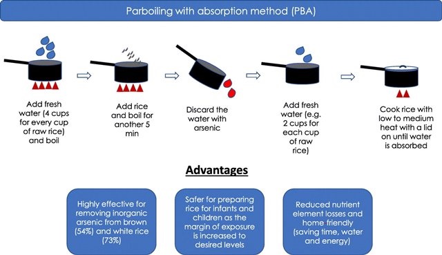
According to their findings, the parboiling with absorption method (PBA) removed the largest amount of arsenic from the rice, while preserving nutrients like phosphorus, potassium, magnesium, zinc, and manganese.

The study was published in Science of The Total Environment.

The method

“With our new method we are able to significantly reduce the arsenic exposure while reducing the loss of key nutrients,” lead author and soil scientist Manoj Menon from the University of Sheffield said in a university statement.

This method, the researchers note, removes over 50% of the naturally occurring arsenic in brown rice, and 74% in white rice, while maintaining micronutrients.

To do this at home, you can do the following, the researchers advise:

  • Bring water to the boil (four cups of fresh water for every cup of raw rice). 
  • Add your rice and boil for another five minutes. 
  • Discard the water (which has now removed much of the arsenic in the rice).
  • Add more fresh water (two cups for each cup of rice). 
  • Finally, cover the rice with a lid, and cook on low to medium heat until the water has been absorbed.

rice

Image credit: Menon et al., Science of The Total Environment, 2020napong detraphiphat

Experiment should be repeated, but a good first step

“We highly recommend this method while preparing rice for infants and children as they are highly vulnerable to arsenic exposure risks,” Menon said.

The researchers say the PBA method also uses less water, energy and cooking time than other cooking methods that can remove the arsenic.

They also encourage additional investigation into using this method in different environments, using different kinds of regional rice types, as well as different levels of water quality – although their experiment is a good first step.

READ | Eating with your eyes – food that looks good doesn’t mean it’s healthier

READ | Popular YouTube channels with kid influencers are promoting unhealthy foods – with lots of views

READ | New device detects heat levels in chillies – so you can avoid the burn

Image: Getty/krisanapong detraphiphat

NEXT ON HEALTH24X

Share Button

Health24.com | OPINION | Unpacking the legal and ethical aspects of South Africa’s Covid-19 track and trace app

  • Tracking and tracing is one of the most important jobs in the pandemic
  • Accuracy a concern, but no evidence to suggest that the app is not working
  • But what about privacy? Apps can be designed to protect privacy rights and to work across borders.

The most effective way to stop the spread of a virus is to prevent contact with everyone who is infected. Those who are infected can be isolated and treated if necessary. To determine who they are, it’s necessary to actively look for and manage cases.

During the Covid-19 pandemic, emerging technologies are being repurposed to help trace whoever has been in contact with an infected person.

Some of these technologies, such as the Global Positioning System (GPS), wi-fi and Bluetooth, are not new. GPS has been used to find accident victims at precise geographic locations. Some of the uses of wi-fi are oxygen monitoring devices, smart beds, access to electronic medical records and real-time access to X-rays and magnetic resonance imaging scans.

Now these tools can also help do one of the most important jobs in the pandemic: track and trace. They allow people some movement and economic activity, with the ability to manage their own risk of being exposed to possible infection or spreading any infection.

The South African government recently introduced a mobile application, COVID Alert SA, to help people notify their close contacts if they are infected.

The app is based on smartphone technology enabled by readily available functions developed by Apple or the Google Exposure Notification System. It uses a phone’s signal to generate a random code. The code is then exchanged with other users of the same app within a two metre radius. These codes are stored on the respective devices for two weeks.

People testing positive for Covid-19 in these two weeks can anonymously alert others by simply clicking a button.

The exchanged and saved codes will, when activated this way, locate and match each other and alert whoever the infected person has been in contact with. This information will allow people to test for Covid-19 and isolate, if necessary, to prevent further infections.

But there are concerns about accuracy, privacy and integration across regions. Questions have also been raised about whether the uptake will be sufficient for the app to function as intended.

In a recent analysis my colleagues and I looked at the legal and ethical aspects of tracing people who might have been in contact with an infected person. Our analysis may provide useful insights when deciding whether to use the app, and help people understand tracking and tracing principles in general.

Key concerns

The first concern is accuracy of the technology. This matters because people’s freedom of movement and ability to go to work or attend school will be affected by an alert.

Cell tower metadata are supplied by electronic communication service providers. The data rely on signal strength and delay times to triangulate the position of a cellular phone. This method often provides inaccurate information because signals are scattered by buildings. There aren’t many towers in rural areas in South Africa, so triangulation may not even be possible there. There’s also a concern that the app might not detect other nearby smartphones accurately.

But there’s no evidence to suggest that the app is not working.

Second, there’s the issue of privacy. The invasive nature of tracking, tracing and surveillance is a real concern.

Before introducing the app, South Africa established a tracing database to serve the same purpose as the current app. The database collected names, identity and cellphone numbers, and the results of people who tested for Covid-19 and their known or suspected contacts. The database had the potential to greatly assist with contact tracing. But it immediately infringed upon the constitutional right to privacy.

To deal with this, the new COVID alert app does not record a person’s geolocation, or any personal or health information. It only uses the location to determine how close another person was and for how long. Even the randomly generated codes cannot be linked to the phone user’s identity. No personal information is recorded or stored anywhere. Hence no privacy rights are at risk of being infringed.

A UK study found that people were more concerned about who was in charge of tracing than they were about their information being stolen. The majority preferred that a health organisation, as opposed to the government, collect and process information. The COVID Alert SA app is officially owned and managed by the South African government.

People also preferred a combination of technology and an old-fashioned human approach to contact tracing.

Public distrust is one of the problems affecting the contact tracing drive in the US. Another is discrepancies in data from different states.

A number of European countries have launched contact tracing apps also using the Google-Apple standard like South Africa. France and Hungary launched different types of apps which will make inter-operability and subsequent tracing across countries in Europe increasingly difficult.

The UK has only very recently joined the countries using the Apple and Google model.

Similarly to the UK and during the early days of the pandemic, China managed to contain the spread of the virus by manual tracing and rapid isolation of positive cases. Now, scientists are calling for technology-based measures to make a long-term response possible.

The winning strategy came from South Korea, whose comprehensive testing and contact tracing was so effective that the country never had to impose a strict lockdown. South Korea used a combination of face-to-face and digital geolocation tracing. It was criticised for infringing on privacy, however. Other countries were more sensitive towards privacy rights.

Effective tool

Digital contact tracing apps can be designed to protect privacy rights and to work across borders.

Research models have found that the more people use these apps, the more transmission of the disease can be reduced and the more scope there is to ease quarantine measures. But the models have also found that the apps are effective even at low uptake levels.

For purposes of managing a pandemic these apps have been tried and tested and evolved to the point where they can do more good than harm if used on a large scale.The Conversation

Dr Marietjie Botes, Post Doctoral Researcher, University of Pretoria

This article is republished from The Conversationunder a Creative Commons license. Read the original article.

Image credit: Unsplash

Share Button

Health24.com | ‘Hidden’ gene discovered in Covid-19 virus could give more insight into its ability to spread

  • Scientists have discovered a ‘hidden’ gene in the genetic code of SARS-CoV-2
  • This overlapping gene, the team said, is unlikely to be detected by the human body’s T-cells
  • Earlier findings have also found mysterious genes, all helpful in the search for treatments and vaccines 

Researchers from the American Museum of Natural History have identified a new “hidden” gene in SARS-CoV-2, the virus that causes Covid-19, and say this may explain why it is so highly infectious.

The discovery of the “overlapping gene”, named ORF3d, could have a significant impact on how we combat the virus, the research team wrote. Overlapping genes (OLGs) are a type of ‘gene within a gene’, effectively concealed in a string of nucleotides, ScienceAlert explains.

“Overlapping genes may be one of an arsenal of ways in which coronaviruses have evolved to replicate efficiently, thwart host immunity, or get themselves transmitted,” said lead author Chase Nelson, a postdoctoral researcher at Academia Sinica in Taiwan and a visiting scientist at the American Museum of Natural History.

“Knowing that overlapping genes exist and how they function may reveal new avenues for coronavirus control, for example through antiviral drugs.”

Their findings were published in the journal eLife.

ORF3d unlikely to be detected by T-cells

According to the team’s findings, the newly discovered gene is present in a previously discovered pangolin coronavirus. This, they say, possibly reveals repeated loss or gain of this gene during the evolution of the new coronavirus, as well as related viruses.

The gene has also shown to elicit a strong antibody response in Covid-19 patients, the researchers said, demonstrating that its protein is manufactured during human infection.

“We don’t yet know its function or if there’s clinical significance,” Nelson said. “But we predict this gene is relatively unlikely to be detected by a T-cell response, in contrast to the antibody response.

“And maybe that has something to do with how the gene was able to arise.”

T-cells are “natural killers”, whose job is to find infected cells in the human body and destroy them, Professor Thomas Scriba, deputy director of immunology and laboratory director at the South African Tuberculosis Vaccine Initiative, University of Cape Town, previously told Health24. 

Mysterious genes explained

Overlapping genes are hard to spot, the researchers explained, as they are virtually hidden from view in the virus’s genome, and therefore easily overlooked.

Most scientific computer programmes are not designed to find them, they said, although OLGs are relatively common in DNA and RNA viruses – partly because RNA viruses (such as SARS-CoV-2) have a high mutation rate.

“Missing overlapping genes puts us in peril of overlooking important aspects of viral biology,” Nelson said.

“In terms of genome size, SARS-CoV-2 and its relatives are among the longest RNA viruses that exist. They are thus perhaps more prone to ‘genomic trickery’ than other RNA viruses.”

Previous discoveries on hidden mutations

Earlier this year, scientists from a separate study also suggested that SARS-CoV-2 exploits a rare, ‘silent’ (hidden) gene mutation in certain people, and said it may provide clarity on why two young and healthy individuals can be so differently affected by the virus, Health24 reported. 

In another breakthrough study, researchers uncovered a second receptor, called neuropilin-1, which may explain why the virus is so highly infectious.

This receptor is widespread in human tissues, they said, and explained that the new coronavirus has a convenient advantage when infecting human cells. These discoveries are all critical in the race to develop a vaccine and treatment for Covid-19.

READ | Leading Covid-19 vaccine candidate 90% effective in Phase 3 trial

READ | The latest news on a leading Covid vaccine candidate is promising, but how optimistic should we be?

READ | Global experts cautiously welcome news of top Covid-19 vaccine candidate’s effectiveness

Image: Getty/Andriy Onufriyenko

Share Button

Is there more to hydration than water?

Mayo Clinic does not endorse companies or products. Advertising revenue supports our not-for-profit mission.

Advertising & Sponsorship

Mayo Clinic Marketplace

Check out these best-sellers and special offers on books and newsletters from Mayo Clinic.

Share Button

COVID-delayed Arctic research cruise yields late-season data

Researchers studying the Bering and Chukchi seas for three weeks in October found no ice and a surprisingly active ecosystem as they added another year’s data to a key climate change record.

The research vessel Norseman II carried scientists from the University of Alaska Fairbanks, the University of Maryland Center for Environmental Science and Clark University.

Maintaining the continuity of long-term observations is crucial as the region is affected by climate change. For example, the researchers collected sediments and small bottom-dwelling animals to help document harmful algal blooms that are becoming more common as Arctic waters warm. The blooms pose a threat to the humans and marine mammals that eat them.

Because of pandemic-related delays, the cruise began on Oct. 2 — a much later start than originally planned. Historically, the Bering and Chukchi sea ecosystem transitioned to lower-level activity as sea ice formed in October.

This year, unseasonably warm ocean temperatures delayed sea ice formation by several weeks. The lack of ice likely allowed the greater biological activity observed by the researchers.

“The recovered data are already showing the effects of oceanic heat that extends further into the fall and early winter,” said Seth Danielson of UAF’s College of Fisheries and Ocean Sciences.

The scientists collected data for several marine science programs monitoring the Pacific Arctic ecosystem.

The Distributed Biological Observatory, led by Jacqueline Grebmeier of UMCES, has been sampling productive hot spots since the late 1980s in U.S. Arctic waters.

The Arctic Marine Biodiversity Observing Network, led by Katrin Iken at UAF’s College of Fisheries and Ocean Sciences, is part of a national network studying how biodiversity and species distributions are changing as a result of climate change in the U.S. Arctic.

The researchers also visited the Chukchi Ecosystem Observatory, a set of highly instrumented oceanographic moorings that monitor the ecosystem year-round. “We only get one chance each year to deploy fresh sensors with new batteries, so this cruise was important to avoid interruptions to the observations,” said Danielson, who leads the project.

“This was a really worthwhile effort that paid off in making biological data available from a part of the year where there have been historically few observations,” said Grebmeier, the cruise’s chief scientist.

To protect communities in the Bering Strait from potential exposure to the COVID-19 virus, the team completed quarantines and multiple tests in Anchorage before the cruise. They traveled by chartered aircraft to Nome and were taken directly to the research vessel, bypassing the passenger terminal.

Everyone aboard also adhered to COVID-19 health and safety mandates from their institutions and followed an isolation and travel plan in accordance with the Port of Nome and the State of Alaska.

Funding for the cruise was provided by the National Oceanic and Atmospheric Administration, the North Pacific Research Board and the National Oceanic Partnership Program, which also includes funding by the Bureau of Ocean Energy Management.

Story Source:

Materials provided by University of Alaska Fairbanks. Note: Content may be edited for style and length.

Share Button