A high number of elderly people on wards and respiratory cases in A&E prompt warnings from staff.


A high number of elderly people on wards and respiratory cases in A&E prompt warnings from staff.

Nearly 19 million adults in the United States take fish oil supplements. These products are rich in omega-3 fatty acids, mainly eicosapentaenoic acid and docosahexaenoic acid, commonly known as EPA and DHA. Many people use them in hopes of lowering inflammation and reducing the risk of chronic diseases.
When it comes to cancer, however, the evidence has been mixed. Some large clinical trials have suggested omega-3 supplements may lower cancer risk, while others have found no benefit or even a possible increase in cancer cases.
A Key Gene in Colon Cancer Protection
Researchers from the University of Michigan and the University of Texas MD Anderson Cancer Center set out to better understand these conflicting results. Their study, published in Cellular and Molecular Gastroenterology and Hepatology, identified a gene called 15-lipoxygenase-1, or ALOX15, as a crucial factor in whether EPA and DHA can help suppress colorectal cancer.
The findings suggest that testing cancer patients for ALOX15 could be important when considering prevention strategies that involve omega-3 supplements.
Surprising Results in Mice
To explore how fish oil affects tumor development, scientists compared mice fed a fish oil enriched diet with mice given a standard diet. Unexpectedly, fish oil increased the number of colon tumors in mice exposed to chemicals that trigger inflammation and speed up tumor growth.
Normally, when EPA and DHA are consumed, the body converts them into compounds known as resolvins. These molecules help reduce chronic inflammation, which plays a major role in cancer development. This conversion process relies on the enzyme ALOX15. However, ALOX15 is often switched off in several types of cancer.
The researchers then examined what happened when mice lacking ALOX15 were given fish oil. In those animals, the absence of ALOX15 led to a rise in colorectal tumors, although the impact varied depending on which omega-3 fatty acid was used.
EPA vs DHA and Different Supplement Forms
Mice fed diets rich in EPA developed fewer tumors than those given DHA. EPA and DHA are available in several forms, including free fatty acids, ethyl esters and triglycerides.
Lovaza, a prescription medication containing the ethyl ester forms of EPA and DHA, is approved by the Food and Drug Administration to treat high triglyceride levels in the blood.
In this study, Lovaza as well as the ethyl ester and free fatty acid forms of EPA reduced both the number and size of tumors, particularly in mice that had active ALOX15. In contrast, DHA variants did not prevent tumor growth in mice lacking ALOX15. When ALOX15 was present, tumor growth was reduced.
“Not all fish oil supplements are the same,” said Imad Shureiqi, professor of internal medicine at the University of Michigan and a member of Rogel Cancer Center.
“It is also important to ask whether the person who is taking the supplement has the required enzymes to metabolize these products to prevent chronic inflammation and subsequently cancer development.”
What This Means for Patients
Although most of the data come from animal studies, the results raise important questions. They suggest that people with colon polyps who do not have active ALOX15 may not receive the same protective benefits from EPA and DHA, making the supplements less effective at slowing tumor growth.
Shureiqi advises patients to speak with their doctors before starting fish oil supplements.
Meanwhile, the research team is developing medications designed to boost ALOX15 levels in cancer cells. Their goal is to enhance the body’s ability to process EPA and DHA, potentially strengthening efforts to prevent colon cancer.

Researchers at Cedars-Sinai have identified a biological repair process that could eventually lead to new treatments for spinal cord injuries, stroke, and neurological diseases such as multiple sclerosis. The findings, published in Nature, reveal an unexpected role for astrocytes, a major support cell in the central nervous system.
“Astrocytes are critical responders to disease and disorders of the central nervous system — the brain and spinal cord,” said neuroscientist Joshua Burda, PhD, assistant professor of Biomedical Sciences and Neurology at Cedars-Sinai and senior author of the study. “We discovered that astrocytes far from the site of an injury actually help drive spinal cord repair. Our research also uncovered a mechanism used by these unique astrocytes to signal the immune system to clean up debris resulting from the injury, which is a critical step in the tissue-healing process.”
The team named these cells “lesion-remote astrocytes,” or LRAs. They also identified several distinct subtypes. For the first time, the study explains how one subtype can detect damage from a distance and respond in ways that support recovery.
How the Spinal Cord Responds to Injury
The spinal cord is a long bundle of nerve tissue that extends from the brain down the back. Its inner region, called gray matter, contains nerve cell bodies along with astrocytes. Surrounding that is white matter, made up of astrocytes and long nerve fibers that carry signals between the brain and the rest of the body. Astrocytes help maintain a stable environment so these signals can travel properly.
When the spinal cord is injured, nerve fibers are torn apart. This can cause paralysis and disrupt sensations such as touch and temperature. The damaged fibers break down into debris. In most tissues, inflammation remains confined to the injured area. In the spinal cord, however, nerve fibers can span long distances, so damage and inflammation can spread well beyond the original injury site.
Lesion-Remote Astrocytes and Immune Cleanup
In experiments involving mice with spinal cord injuries, researchers found that LRAs play a key role in promoting repair. They also found strong signs that the same process occurs in spinal cord tissue from human patients.
One LRA subtype produces a protein called CCN1. This molecule sends signals to immune cells known as microglia.
“One function of microglia is to serve as chief garbage collectors in the central nervous system,” Burda said. “After tissue damage, they eat up pieces of nerve fiber debris — which are very fatty and can cause them to get a kind of indigestion. Our experiments showed that astrocyte CCN1 signals the microglia to change their metabolism so they can better digest all that fat.”
According to Burda, this improved debris removal may help explain why some patients experience partial, spontaneous recovery after spinal cord injury. When researchers eliminated astrocyte-derived CCN1, healing was significantly reduced.
“If we remove astrocyte CCN1, the microglia eat, but they don’t digest. They call in more microglia, which also eat but don’t digest,” Burda said. “Big clusters of debris-filled microglia form, heightening inflammation up and down the spinal cord. And when that happens, the tissue doesn’t repair as well.”
Implications for Multiple Sclerosis and Brain Injury
When scientists examined spinal cord samples from people with multiple sclerosis, they observed the same CCN1-related repair process. Burda noted that these basic repair principles may apply broadly to injuries affecting either the brain or spinal cord.
“The role of astrocytes in central nervous system healing is remarkably understudied,” said David Underhill, PhD, chair of the Department of Biomedical Sciences. “This work strongly suggests that lesion-remote astrocytes offer a viable path for limiting chronic inflammation, enhancing functionally meaningful regeneration, and promoting neurological recovery after brain and spinal cord injury and in disease.”
Burda is now working to develop strategies that harness the CCN1 pathway to improve spinal cord healing. His team is also studying how astrocyte CCN1 may influence inflammatory neurodegenerative diseases and aging.
Additional Cedars-Sinai authors include Sarah McCallum, Keshav B. Suresh, Timothy S. Islam, Manish K. Tripathi, Ann W. Saustad, Oksana Shelest, Aditya Patil, David Lee, Brandon Kwon, Katherine Leitholf, Inga Yenokian, Sophia E. Shaka, Jasmine Plummer, Vinicius F. Calsavara, and Simon R.V. Knott.
Other authors include Connor H. Beveridge, Palak Manchandra, Caitlin E. Randolph, Gordon P. Meares, Ranjan Dutta, Riki Kawaguchi, and Gaurav Chopra.
Funding: This work was supported by: the US National Institutes of Health (NIH) 5R01NS128094, R00NS105915, K99NS105915 (to J.E.B.), F31NS129372 (to K.S.), K99AG084864 (S.M.) R35 NS097303 and R01 NS123532 (RD), R01MH128866, U18TR004146, P30 CA023168 and ASPIRE Challenge and Reduction-to-Practice award (to G.C.); the Paralyzed Veterans Research Foundation of America (to J.E.B.); Wings for Life (to J.E.B.); Cedars-Sinai Center for Neuroscience and Medicine Postdoctoral Fellowship (to S.M.); American Academy of Neurology Neuroscience Research Fellowship (to S.M.); California Institute for Regenerative Medicine Postdoctoral Scholarship (to S.M.); The United States Department of Defense USAMRAA award W81XWH2010665 through the Peer Reviewed Alzheimer’s Research Program (to G.C.); The Arnold O. Beckman Postdoctoral Fellowship (to C.E.R.); The Purdue University Center for Cancer Research funded by NIH grant P30 CA023168 is also acknowledged.
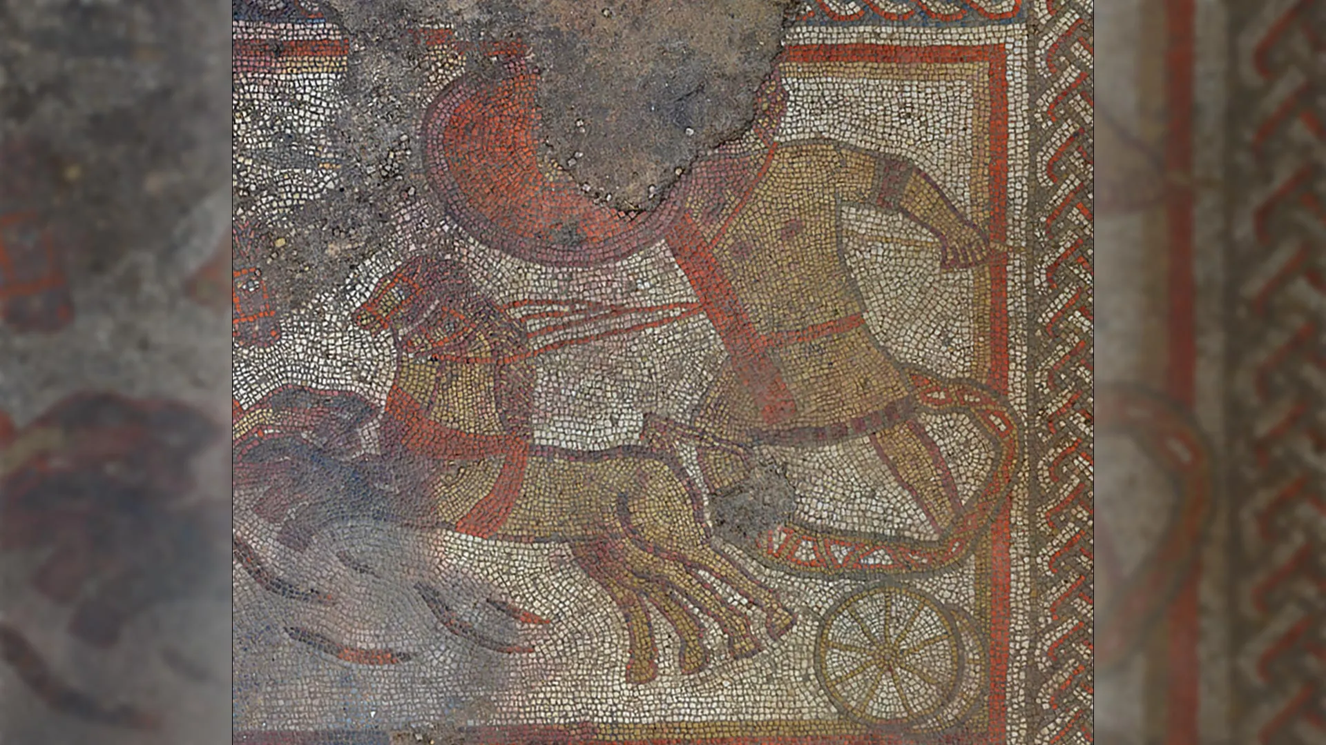
Researchers studying what has been called one of the most important mosaics ever found in the UK say it portrays a rarely told version of the Trojan War that had largely faded from history.
A new investigation by the University of Leicester explains why the well known Ketton mosaic in Rutland — considered one of the most remarkable Roman era discoveries in Britain in the past century — does not illustrate scenes from Homer’s Iliad, as scholars first believed. Instead, the artwork reflects a different telling of the Trojan War that was made famous by the Greek playwright Aeschylus. That dramatic version has not survived in full and is considered lost.
The findings also show that the mosaic incorporates decorative motifs that had been circulating around the Mediterranean for centuries. This suggests that artists working in Roman Britain were closely connected to broader artistic traditions across the classical world.
Discovery During Lockdown
The Ketton mosaic came to light in 2020 during the COVID-19 lockdown, when local resident Jim Irvine spotted it on his family farm. The discovery led to a major excavation by University of Leicester Archaeological Services (ULAS), funded by Historic England. The mosaic and the surrounding Roman villa have since been granted Scheduled Monument status because of their national significance. Historic England and ULAS carried out joint excavations in 2021 and 2022 and are now preparing the full publication of their findings.
The mosaic features three dramatic moments involving the Greek warrior Achilles and the Trojan prince Hector: their fierce duel, Achilles dragging Hector’s body, and the later scene in which King Priam ransoms his son’s body, which is shown being weighed in gold.
The Trojan War, most famously described in Homer’s epic poem the Iliad, tells of a legendary ten year conflict between Greek forces and the city of Troy, ruled by King Priam. According to myth, the war began after Helen of Sparta was taken to Troy.
Aeschylus and a Forgotten Greek Tragedy
Detailed analysis now indicates that the Ketton mosaic does not follow Homer’s narrative. Instead, it closely matches Phrygians, a lesser known tragedy written by the Athenian playwright Aeschylus. While Romans were familiar with multiple versions of the Trojan War story, choosing this more unusual interpretation would have set the villa’s owner apart and signaled cultural sophistication.
The study also highlights how the mosaic blends long established artistic designs from across the Mediterranean.
Dr. Jane Masséglia, lead author of the study and Associate Professor in Ancient History at the University of Leicester, said: “In the Ketton Mosaic, not only have we got scenes telling the Aeschylus version of the story, but the top panel is actually based on a design used on a Greek pot that dates from the time of Aeschylus, 800 years before the mosaic was laid. Once I’d noticed the use of standard patterns in one panel, I found other parts of the mosaic were based on designs that we can see in much older silverware, coins and pottery, from Greece, Turkey, and Gaul.
“Romano-British craftspeople weren’t isolated from the rest of the ancient world, but were part of this wider network of trades passing their pattern catalogues down the generations. At Ketton, we’ve got Roman British craftsmanship but a Mediterranean heritage of design.”
Roman Britain and the Wider Ancient World
Jim Irvine, who first uncovered the mosaic in 2020, said: “Jane’s detailed research into the Rutland mosaic imagery reveals a level of cultural integration across the Roman world that we’re only just beginning to appreciate. It’s a fascinating and important development that suggests Roman Britain may have been far more cosmopolitan than we often imagine. The new paper is a suspenseful and thrilling narrative in its own right which deserves recognition.”
Rachel Cubitt, Post-Excavation Coordinator at Historic England, added: “Working in collaboration with the University of Leicester brings an added dimension to investigations at the Ketton villa site. This fascinating new research offers a more nuanced picture of the interests and influences of those who may have lived there, and of people living across Roman Britain at this time.”
Hella Eckhardt, Professor of Roman Archaeology at the University of Reading, who was not involved in the study, said: “This is an exciting piece of research, untangling the ways in which the stories of the Greek heroes Achilles and Hector were transmitted not just through texts but through a repertoire of images created by artists working in all sorts of materials, from pottery and silverware to paintings and mosaics.”
Side effects of a common Parkinson’s medications had devastating consequences on one family, BBC hears.
The plans are subject to a 12-week public consultation which starts on Friday.
Scientists have discovered how to make people less selfish – slightly and temporarily – by stimulating two areas of the brain.
Unions angry, but government says it demonstrates its commitment to workforce.
Scientists at the Yong Loo Lin School of Medicine at the National University of Singapore have identified a protein that may help restore the brain’s ability to produce new cells as it ages. Their findings, published in Science Advances, point to a transcription factor called cyclin D-binding myb-like transcription factor 1 (DMTF1) as a central regulator of neural stem cell activity in older brains. Transcription factors are proteins that control how genes are turned on or off in specific cells.
Neural stem cells are responsible for generating new neurons, which play an essential role in learning and memory. As people age, these stem cells gradually lose their ability to renew themselves, contributing to cognitive decline.
Investigating DMTF1 in Aging Brain Cells
The study was led by Assistant Professor Ong Sek Tong Derrick, with Dr. Liang Yajing as first author, from the Department of Physiology and the Healthy Longevity Translational Research Programme at NUS Medicine. The team set out to uncover the biological changes that cause neural stem cells to weaken over time, with the goal of identifying targets for future therapies aimed at slowing neurological aging.
To understand how DMTF1 functions, the researchers examined neural stem cells derived from humans and from laboratory models designed to mimic premature aging. They used genome binding and transcriptome analyses to map how DMTF1 influences gene activity. A key focus was how this protein interacts with stem cells affected by telomere dysfunction. Telomeres are the protective ends of chromosomes that gradually shorten each time a cell divides. This shortening is widely recognized as a marker of aging.
Restoring Regeneration in Aged Stem Cells
The team found that levels of DMTF1 were significantly reduced in “aged” neural stem cells. When they restored DMTF1 expression, the cells regained their ability to regenerate. This suggests that DMTF1 could serve as a promising therapeutic target for restoring stem cell function in the aging brain.
Further analysis revealed how DMTF1 exerts its effects. The protein regulates helper genes (Arid2 and Ss18) that loosen tightly packed DNA, allowing growth-related genes to become active. Without these helper genes, neural stem cells cannot effectively renew themselves.
“Impaired neural stem cell regeneration has long been associated with neurological aging. Inadequate neural stem cell regeneration inhibits the formation of new cells needed to support learning and memory functions. While studies have found that defective neural stem cell regeneration can be partially restored, its underlying mechanisms remain poorly understood,” said Asst Prof Ong. “Understanding the mechanisms for neural stem cell regeneration provides a stronger foundation for studying age-related cognitive decline.”
Potential Therapies to Slow Brain Aging
The findings indicate that strategies designed to increase DMTF1 levels or enhance its activity could potentially reverse or delay the decline in neural stem cell function linked to aging.
Although the current results are based largely on in vitro experiments, the researchers plan to investigate whether boosting DMTF1 can increase neural stem cell numbers and improve learning and memory in conditions involving telomere shortening and natural aging, without raising the risk of brain tumours. Over the long term, the team hopes to identify small molecules capable of safely stimulating DMTF1 activity to rejuvenate aging neural stem cells.
“Our findings suggest that DMTF1 can contribute to neural stem cell multiplication in neurological aging,” Dr. Liang said. “While our study is in its infancy, the findings provide a framework for understanding how aging-associated molecular changes affect neural stem cell behavior, and may ultimately guide the development of successful therapeutics.”

Picture Europe tens of thousands of years ago. Thick forests covered much of the land. Herds of elephants, bison, and aurochs roamed freely. Small bands of humans moved through this world carrying fire and spears.
New research suggests those early people changed their surroundings far more than scientists once believed.
Computer Models Reveal Early Human Impact
An international team led in part by researchers at Aarhus University used advanced computer simulations to examine how climate, large animals, natural fires, and humans shaped European vegetation during two past warm periods. The team then compared those simulations with extensive fossil pollen data from the same eras. By matching the models with real world evidence preserved in pollen, they were able to estimate how much each factor influenced plant cover.
The results point to a clear conclusion. Both Neanderthals and later Mesolithic hunter-gatherers significantly altered vegetation patterns across Europe, long before agriculture began.
“The study paints a new picture of the past,” says Jens-Christian Svenning, professor of biology at Aarhus University. The project involved experts in archaeology, geology, and ecology from The Netherlands, Denmark, France, and UK.
“It became clear to us that climate change, large herbivores and natural fires alone could not explain the pollen data results. Factoring humans into the equation — and the effects of human-induced fires and hunting — resulted in a much better match,” says Jens-Christian Svenning.
The findings were recently published in PLOS One.
Humans and the Decline of Megafauna
The researchers focused on two specific warm intervals in European history.
The first was the Last Interglacial period around 125,000-116,000 years ago, when Neanderthals were the only humans living in Europe. The second was the Early Holocene, 12,000-8,000 years ago, just after the last Ice Age, when Mesolithic hunter-gatherers from our own species, Homo sapiens, inhabited the region.
During the Last Interglacial, Europe supported a wide range of megafauna. Elephants and rhinoceroses lived alongside bison, aurochs, horses, and deer.
By the Mesolithic period, that picture had changed. Many of the largest animals had vanished or their numbers had dropped sharply. This reflected the broader wave of megafauna losses that followed the global spread of Homo sapiens.
A New View of Prehistoric Europe
“Our simulations show that Mesolithic hunter-gatherers could have influenced up to 47% of the distribution of plant types. The Neanderthal effect was smaller, but still measurable — approximately 6% for plant type distribution and 14% for vegetation openness,” says Anastasia Nikulina.
Human influence showed up in two main ways. One was the use of fire, which burned trees and shrubs. The other was hunting large herbivores, a factor that has often been overlooked.
“The Neanderthals did not hold back from hunting and killing even giant elephants. And here we’re talking about animals weighing up to 13 tonnes. Hunting also had a strong indirect effect: fewer grazing animals meant more overgrowth and thus more closed vegetation. However, the effect was limited, because the Neanderthals were so few that they did not do eliminate the large animals or their ecological role — unlike Homo sapiens in later times,” says Jens-Christian Svenning.
According to Nikulina and Svenning, these findings challenge the idea that Europe was an untouched wilderness before farming began.
“The Neanderthals and the Mesolithic hunter-gatherers were active co-creators of Europe’s ecosystems,” says Jens-Christian Svenning. “The study is consistent with both ethnographic studies of contemporary hunter-gatherers and archaeological finds, but goes a step further by documenting how extensive human influence may have been tens of thousands of years ago — that is, before humans started farming the land,” elaborates Anastasia Nikulina.
AI Simulations and Interdisciplinary Research
Nikulina emphasizes that the project brought together multiple fields, including ecology, archaeology and palynology (knowledge about pollen). The team also developed detailed computer models to simulate ancient ecosystems.
“This is the first simulation to quantify how Neanderthals and Mesolithic hunter-gatherers may have shaped European landscapes. Our approach has two key strengths: it brings together an unusually large set of new spatial data spanning the whole continent over thousands of years, and it couples the simulation with an optimization algorithm from AI. That let us run a large number of scenarios and identify the most possible outcomes,” says Anastasia Nikulina.
Svenning adds that the modeling made one thing clear.
“The computer modeling made it clear to us that climate change, the large herbivores such as elephants, bison and deer, and natural wildfires alone cannot explain the changes seen in ancient pollen data. To understand the vegetation at that time, we must also take human impacts into account — both direct and indirect. Even without fire, hunter-gatherers changed the landscape simply because their hunting of large animals made the vegetation denser,” says Jens-Christian Svenning.
Even with these advances, gaps remain in our knowledge about how early humans influenced their environments.
Nikulina and Svenning note that similar simulations could be applied to other regions and time periods. North and South America and Australia are especially intriguing because they were not inhabited by earlier hominin species before Homo sapiens. That makes it possible to compare more recent landscapes with and without human presence.
“And although the large models paint a broad picture, detailed local studies are absolutely essential to improve our understanding of the way humans shaped the landscape in prehistoric times,” says Jens-Christian Svenning.
