Breakthrough brain discovery reveals a natural way to relieve pain

  • Scientists have discovered that the human brain has its own built-in pain map, activating different regions when easing pain in the face, arms, or legs.
  • Placebo pain relief only works in the exact area where the brain expects it to happen.
  • Understanding this system could lead to safer, more precise treatments that target pain exactly where it occurs.

Mapping the Brain’s Hidden System for Pain

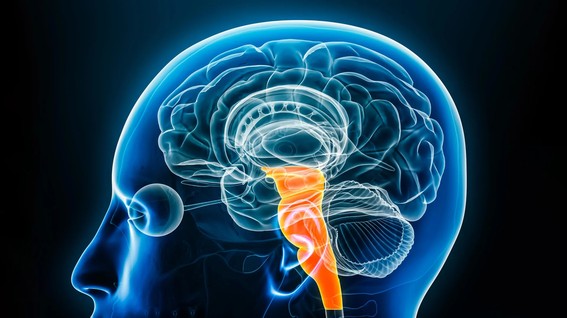
Researchers at the University of Sydney have revealed a brainstem network that manages pain differently depending on where it occurs in the body. Using placebo pain relief, they discovered a map-like system that fine-tunes pain control for specific regions, such as the face, arms, or legs. The findings could lead to safer and more precise pain therapies that avoid the risks of opioid-based treatments.

The brainstem functions as the main communication pathway between the brain and spinal cord, directing signals that control thought, sensation, and survival responses. It also produces most of the brain’s vital neurochemicals, making it a central hub for regulating both physical and emotional states.

Published in Science, the study used 7-Tesla functional magnetic resonance imaging (fMRI) (one of the most advanced brain scanners available, with only two in Australia) to identify how two major regions of the brainstem coordinate pain relief through placebo responses.

Dr. Lewis Crawford, lead author and research fellow at the School of Medical Sciences and the Brain and Mind Centre, explained, “This is the first time we’ve seen such a precise and detailed pain map in the human brainstem, showing us that it tailors pain relief to the specific part of the body that’s experiencing it.”

This breakthrough builds on decades of research led by co-author Professor Kevin Keay, Deputy Head of the School of Medical Sciences, who has long studied the brain’s role in pain regulation.

How the Placebo Effect Reveals the Brain’s Pain Control

To explore how the brain organizes pain relief, researchers tested 93 healthy volunteers by applying heat to different parts of their bodies. A placebo cream was used on some areas, but scientists secretly lowered the temperature to convince participants that the cream was reducing pain.

Each participant’s heat level was personalized to reach a moderate level of discomfort, based on a scale from 0 (no pain) to 100 (worst pain imaginable), typically between 40 and 50 degrees Celsius.

When the same heat stimulus was later reapplied, participants continued to feel less pain in the areas where the placebo cream had been used, even though the temperature was no longer reduced. About 61 percent reported this effect, a strong indication of genuine placebo-driven pain relief.

Dr. Crawford noted, “We found that upper parts of the brainstem were more active when relieving facial pain, while lower regions were engaged for arm or leg pain.”

Pinpointing the Brain’s Pain-Relief Centers

Two major brainstem regions, the periaqueductal grey (PAG) and the rostral ventromedial medulla (RVM), were identified as central to this system. Each showed distinct patterns of activity depending on where the pain occurred. Upper sections of the PAG and RVM responded to facial pain, while lower sections activated for pain in the limbs.

According to Dr. Crawford, “The brain’s natural pain relief system is more nuanced than we thought. Essentially, it has a built-in system to control pain in specific areas. It’s not just turning pain off everywhere; but working in a highly coordinated, anatomically precise system.”

A Blueprint for Targeted Pain Therapies

Understanding which brainstem areas are linked to different parts of the body may open new avenues for developing non-invasive therapies that reduce pain without widespread side effects.

“We now have a blueprint for how the brain controls pain in a spatially organized way,” said Professor Luke Henderson, senior author and Professor in the School of Medical Sciences and the Brain and Mind Centre. “This could help us design more effective and personalized treatments, especially for people with chronic pain in a specific area of their body.”

The study also challenges long-held assumptions about how placebo pain relief works. Instead of relying on the brain’s opioid system, experts say a different part of the brainstem — the lateral PAG — is not only responsible but works without using opioids and could instead be linked to cannabinoid activity.

“Opioid-based pain relief typically activates central areas of the brain and can affect the whole body, whereas the cannabinoid circuit that we identified appears to operate in more targeted regions of the brainstem,” said Dr. Crawford. “This supports the idea that cannabinoids may play a role in localized, non-opioid pain control.”

“Knowing exactly where pain relief is happening in the brain means we can target that area or assess whether a drug is working in the right place,” said Dr. Crawford. “This could lead to more precise treatments for chronic pain that don’t rely on opioids and work exactly where the brain expects pain relief to occur — a huge step forward for pain management.”


Share Button

Think melatonin is safe? New research reveals a hidden heart risk

  • A large review of health data from more than 130,000 adults with insomnia found that people who took melatonin for a year or longer were more likely to develop heart failure, be hospitalized for the condition, or die from any cause compared to those who didn’t take the supplement.
  • While the study cannot prove that melatonin directly causes these outcomes, the strong association raises important safety questions about long-term use of this popular sleep aid. Researchers emphasize that more studies are needed to fully understand melatonin’s impact on heart health and ensure it can be used safely.

Long-Term Melatonin Use Linked to Higher Heart Failure Risk

People who regularly take melatonin to improve sleep may face serious health risks. A preliminary study presented at the American Heart Association’s Scientific Sessions 2025 found that adults with chronic insomnia who used melatonin for a year or longer were more likely to develop heart failure, be hospitalized for heart failure, and die from any cause than those who did not take the supplement. The findings will be discussed at the AHA’s annual meeting, taking place Nov. 7-10 in New Orleans, a leading international event for cardiovascular science and clinical research updates.

Melatonin is a hormone produced by the pineal gland that regulates the body’s sleep-wake cycle. Its levels naturally rise in the dark and drop during daylight hours. Synthetic melatonin, which is chemically identical to the natural hormone, is widely used to treat insomnia (difficulty falling and/or staying asleep) and jet lag. In many countries, including the U.S., melatonin supplements can be purchased over the counter. However, because they are not regulated in the U.S., products can differ widely in purity and dosage.

How the Study Was Conducted

Researchers divided participants into two groups based on their medical records. Those who had taken melatonin for at least one year were classified in the “melatonin group,” while individuals with no record of melatonin use were placed in the “non-melatonin group.”

“Melatonin supplements may not be as harmless as commonly assumed. If our study is confirmed, this could affect how doctors counsel patients about sleep aids,” said Ekenedilichukwu Nnadi, M.D., lead author of the study and chief resident in internal medicine at SUNY Downstate/Kings County Primary Care in Brooklyn, New York.

Investigating Heart Failure and Sleep Aid Safety

Although melatonin is marketed as a safe and natural sleep remedy, little evidence exists on its long-term cardiovascular effects. The research team wanted to know whether long-term use could influence heart failure risk in people with chronic insomnia. According to the American Heart Association’s 2025 Heart Disease and Stroke Statistics, heart failure occurs when the heart cannot pump enough oxygen-rich blood to sustain the body’s organs. The condition affects about 6.7 million U.S. adults.

To explore this question, scientists used data from the TriNetX Global Research Network, an international database of de-identified medical records. They reviewed five years of data on adults diagnosed with chronic insomnia who had documented melatonin use for more than a year. Each was matched with another person who also had insomnia but had never used melatonin. Individuals with a previous diagnosis of heart failure or who had been prescribed other sleep medications were excluded.

The main analysis found:

  • Among adults with insomnia, those whose electronic health records indicated long-term melatonin use (12 months or more) had about a 90% higher chance of incident heart failure over 5 years compared with matched non-users (4.6% vs. 2.7%, respectively).
  • There was a similar result (82% higher) when researchers analyzed people who had at least 2 melatonin prescriptions filled at least 90 days apart. (Melatonin is only available by prescription in the United Kingdom.)

A secondary analysis found:

  • Participants taking melatonin were nearly 3.5 times as likely to be hospitalized for heart failure when compared to those not taking melatonin (19.0% vs. 6.6%, respectively).
  • Participants in the melatonin group were nearly twice as likely to die from any cause than those in the non-melatonin group (7.8% vs. 4.3%, respectively) over the 5-year period.

“Melatonin supplements are widely thought of as a safe and ‘natural’ option to support better sleep, so it was striking to see such consistent and significant increases in serious health outcomes, even after balancing for many other risk factors,” Nnadi said.

Expert Reactions and Caution From Sleep Researchers

“I’m surprised that physicians would prescribe melatonin for insomnia and have patients use it for more than 365 days, since melatonin, at least in the U.S., is not indicated for the treatment of insomnia. In the U.S., melatonin can be taken as an over-the-counter supplement and people should be aware that it should not be taken chronically without a proper indication,” said Marie-Pierre St-Onge, Ph.D., C.C.S.H., FAHA, chair of the writing group for the American Heart Association’s 2025 scientific statement, Multidimensional Sleep Health: Definitions and Implications for Cardiometabolic Health. St-Onge, who was not involved in this study, is a professor of nutritional medicine in the division of general medicine and director of the Center of Excellence for Sleep & Circadian Research in the department of medicine at Columbia University Irving Medical Center in New York City.

The study has several limitations. First, the database includes countries that require a prescription for melatonin (such as the United Kingdom) and countries that don’t (such as the United States), and patient locations were not part of the de-identified data available to the researchers. Since melatonin use in the study was based only on those identified from medication entries in the electronic health record, everyone taking it as an over-the-counter supplement in the U.S. or other countries that don’t require a prescription would have been in the non-melatonin group; therefore, the analyses may not accurately reflect this. Hospitalization figures were also higher than those for initial diagnosis of heart failure because a range of related diagnostic codes may be entered for the hospitalization, and they may not always include the code for a new diagnosis of heart failure. The researchers also lacked information on the severity of insomnia and the presence of other psychiatric disorders.

“Worse insomnia, depression/anxiety or the use of other sleep-enhancing medicines might be linked to both melatonin use and heart risk,” Nnadi said. “Also, while the association we found raises safety concerns about the widely used supplement, our study cannot prove a direct cause-and-effect relationship. This means more research is needed to test melatonin’s safety for the heart.”

Study details, background and design:

  • The study included 130,828 adults (average age of 55.7 years; 61.4% women) diagnosed with insomnia.
  • The study data was from TriNetX, established in 2013, a growing global network of real-world, de-identified patient data available for research.
  • 65,414 participants had been prescribed melatonin at least once and reported taking it for at least a year.
  • A second group of people were examined for comparison (control group) — those who had never been prescribed melatonin and were matched to the group taking melatonin on 40 factors including demographic information, health conditions and medications.
  • Participants were excluded if they had already been diagnosed with heart failure or had been prescribed other types of sleeping pills such as benzodiazepines.
  • The melatonin and control groups were matched for age, sex, race/ethnicity, heart and nervous system diseases, medications for heart and nervous system diseases, blood pressure and body mass index. Researchers looked at electronic medical records from the five years after the matching date.
  • For the main findings, records were searched for codes related to an initial diagnosis of heart failure. Secondary findings included codes for hospitalization related to heart failure or death.
  • Following the initial analyses, researchers validated the credibility of their findings by conducting a sensitivity analysis. This involved slightly changing the criteria: they required participants in the melatonin group to have filled at least two melatonin prescriptions that were at least 90 days apart. This adjustment aimed to determine whether the extended duration of confirmed melatonin prescriptions influenced the outcomes.

Note: The study featured in this article is a research abstract. Abstracts presented at American Heart Association’s scientific meetings are not peer-reviewed, and the findings are considered preliminary until published as full manuscripts in a peer-reviewed scientific journal.

Share Button

Calls for legal right to paid leave for IVF treatment

Campaigners say IVF treatment should be less stigmatised in the workplace and come with legal entitlements to time off

Share Button

What Each Ripeness Level Of Your Banana Actually Means For Your Health

Here’s something you may not have thought about: For some foods (in this case we’re talking about bananas), the nutritional effects on your health can vary depending on the ripeness level when you eat it.

As a banana ripens, its nutritional content — the amount of sugar, starch and vitamins — changes. Meaning, if you need more sugar before a workout, or less sugar because of diabetes, you may want to eat your banana at a certain ripeness level.

Ahead, registered dieticians share the various ripeness levels, what a banana looks like at each stage, and the health conditions or goals that are best addressed at each level of ripeness.

Underripe

An underripe banana can help you feel full longer.

Dorin Musat / 500px via Getty Images

An underripe banana can help you feel full longer.

Bananas at this stage look green; they’re firm and difficult to peel.

“This stage has the highest resistant starch and low sugar content,” said Avery Zenker, a registered dietitian at MyCrohnsAndColitisTeam.

The benefits of resistant starch are many, including helping to feed healthy gut bacteria that reduce inflammation and keeping blood sugar levels stable, according to Zenker. It’s also digested more slowly, which can keep you full for longer.

“Resistant starch acts very similarly to fibre, which means it’s fermented in the gut (so it fuels your good gut bacteria) and it can be helpful for blood sugar control,” added registered dietitian Amanda Sauceda, a nutrition lecturer at California State University, Long Beach.

While good gut bacteria and blood sugar management are generally important for your health, they may apply to some individuals more than others.

“This level of ripeness is ideal for people who are trying to control blood glucose levels, such as those with insulin resistance as seen with pre-diabetes, Type 2 diabetes and other metabolic conditions,” Zenker said. “Unripe bananas can also be beneficial for anyone looking to improve gut health by improving microbiome balance.” The latter may be especially important for people who are trying to manage digestive issues like irritable bowel syndrome.

“If you want to up the health benefits of your green banana, consider pairing it with some peanut butter,” Sauceda added. “You get a little protein and healthy fats, which help for a better blood sugar response and are good for the gut.”

Some people may want to avoid an underripe banana, however. Zenker said athletes may fall into that category since slow digestion can cause stomach heaviness. People who are sensitive to resistant starch and experience gastrointestinal discomfort after may want a different ripeness level, too.

Barely Ripe

Barely ripe bananas still have a little green near the ends.

mrs via Getty Images

Barely ripe bananas still have a little green near the ends.

Bananas at this stage are mostly yellow with some green at the ends. While slightly softer, they’re still firm. This ripeness level suggests the banana is still high in fibre and low in sugar, but not to the same extent as underripe bananas.

“The resistant starches have started converting into simple sugars,” Zenker said. “Their mineral levels remain stable, particularly potassium and magnesium.”

She recommended a barely ripe banana to people who want the digestive benefits of an underripe banana without that chalky taste, as well as those who need steady energy throughout the day without major effects on their blood sugar. That means, similar to underripe bananas, barely ripe ones are great for people with insulin resistance, prediabetes, diabetes, metabolic syndrome or digestive conditions.

“They can also be ideal for peri- or post-menopausal women due to the resistant starch content that helps combat the effects of insulin resistance and supports gut health as hormonal changes occur,” Zenker added.

Ripe

A fully ripe banana has no green showing on the ends.

Yaorusheng via Getty Images

A fully ripe banana has no green showing on the ends.

Bananas at this stage are fully yellow and soft, but not mushy. They’re easy to peel and have a sweet smell.

While ripe bananas are listed separately from barely ripe ones here, it’s important to note that not all experts find the differences noteworthy.

“Nutrition-wise, these types of bananas are pretty much the same,” Sauceda said. “In fact, the USDA nutrient database categorizes these two types of bananas together.”

With that said, if we’re going to make distinctions, Zenker shared what they are: In ripe bananas, starches have mostly converted into natural sugars, fiber decreases, and sugar and antioxidant content increase. Vitamins and minerals hit their peak.

Sauceda added more noteworthy benefits. “One banana has 8% of your daily value for potassium, which is a mineral many people don’t get enough of,” she said. “This mineral is important for blood pressure and muscle contraction. It also meets 8% of your daily value for magnesium, which is important for bone health.”

Zenker encouraged a ripe banana for people who are about to work out, want quick and convenient whole-food energy, or are looking to increase potassium intake (perhaps to manage high blood pressure or other cardiovascular issues), as well as for kids who want an easy-to-eat, sugary food. Ripe bananas are also a good in-between for people who want fibre without the digestion-related concerns.

Very Ripe

Two very ripe bananas, indicated by brown spots on the peel.

Stefania Pelfini la Waziya via Getty Images

Two very ripe bananas, indicated by brown spots on the peel.

Bananas at this stage are soft, have brown spots and emit a strong aroma.

“Sugar content is near its peak, and fibre continues to decrease,” Zenker said.

That’s great for people who need quick energy, have sensitive digestive symptoms, have low appetite or want a sweet treat, Zenker said. Individuals who are trying to manage their blood sugar may want to avoid very ripe bananas, however, since they’re higher in sugar.

Overripe

An overripe banana looks brown or black and is usually mushy.

Karl Tapales via Getty Images

An overripe banana looks brown or black and is usually mushy.

Bananas at this stage look brown or black and are soft, even mushy.

They’re highest in sugar and antioxidants, lowest in fibre, losing their vitamin C content and maintaining their potassium content, according to Zenker.

As far as other vitamins go, some are increasing while others are decreasing. “For example, a ripe banana has 14 milligrams of vitamin C but an overripe one has 10 milligrams,” Sauceda said. “Interestingly, overripe bananas have a bit more folate than ripe bananas.”

So again, there are pros and cons, and certain populations and situations for which it’s best suited.

“This stage is best for baking and freezing (banana bread, cookies, smoothies) rather than as a raw snack,” Zenker said. “It’s gentle on digestion and good for anyone needing fast calories, such as athletes, [but] not as ideal for people with diabetes or those seeking fibre or nutrient density.”

Sauceda is a fan of the smoothie option. “You don’t need to add as much banana when you use an overripe one for your smoothie, which can mean less added sugars,” she said.

As evidenced here, the banana to pick at the grocery store isn’t necessarily just the one you like the taste or colour of. Yes, we know this concept is a bit bananas, literally — and it’s true.

At the same time, we can’t blame you for going by the taste metric. Plus, bananas are nutritious whenever you eat them. “Overall, bananas are a great fruit whether you eat them green or ripe,” Sauceda said.

Share Button

Maldives bans smoking for younger generations

Anyone born on or after 1 January 2007 will be banned from using, buying or selling tobacco products in the archipelago.

Share Button

Astronomers capture a violent super-eruption from a young sun

Although we rarely notice from Earth, the Sun is continuously hurling enormous clouds of charged plasma into space. These events, known as coronal mass ejections (CMEs), often occur alongside sudden bursts of light called solar flares. When particularly strong, CMEs can stretch far enough to disturb Earth’s magnetic field, producing dazzling auroras and sometimes triggering geomagnetic storms that disrupt satellites or even power grids.

Scientists believe that billions of years ago, when the Sun and Earth were both young, solar activity was far more intense than it is today. Powerful CMEs during that period may have influenced the conditions that allowed life to emerge and evolve. Studies of young Sun-like stars — used as stand-ins for our own star’s early years — show that these stars often unleash flares far stronger than any recorded from the modern Sun.

Reconstructing Ancient Solar Explosions

Massive eruptions from the early Sun likely had dramatic effects on the atmospheres of Earth, Mars, and Venus. Yet researchers still do not fully understand how closely these stellar outbursts resemble today’s CMEs. While scientists have recently observed cooler plasma components of CMEs from the ground, detecting the fast-moving, high-energy events expected in the past has proven much more difficult.

To explore this question, an international research team led by Kosuke Namekata of Kyoto University set out to determine whether young Sun-like stars generate CMEs similar to those of our own Sun.

“What inspired us most was the long-standing mystery of how the young Sun’s violent activity influenced the nascent Earth,” says Namekata. “By combining space- and ground-based facilities across Japan, Korea, and the United States, we were able to reconstruct what may have happened billions of years ago in our own solar system.”

The researchers conducted simultaneous ultraviolet observations with the Hubble Space Telescope and optical observations from ground-based telescopes in Japan and Korea. Their subject was the young Sun-like star EK Draconis. Hubble measured ultraviolet light from extremely hot plasma, while the ground-based observatories tracked cooler hydrogen gas through the Hα line. This coordinated, multi-wavelength approach enabled the team to capture both the hot and cool parts of a CME as it unfolded.

Evidence of a Multi-Temperature Solar Eruption

The observations revealed the first-ever evidence of a multi-temperature CME from EK Draconis. The team discovered that plasma heated to about 100,000 degrees Kelvin was expelled at speeds of 300 to 550 kilometers per second (~670,000 to 1,230,000 miles per hour). Roughly ten minutes later, cooler gas around 10,000 degrees was launched at about 70 kilometers per second (~160,000 miles per hour). The high-temperature plasma carried significantly more energy, indicating that frequent and powerful CMEs in the past could have produced strong shocks and energetic particles capable of reshaping or stripping early planetary atmospheres.

Other studies support the idea that energetic solar events and their resulting particles may have triggered chemical reactions that produced biomolecules and greenhouse gases — key ingredients for sustaining life. This finding therefore deepens our understanding of how solar activity may have created the environmental conditions necessary for life to appear on early Earth, and possibly on other planets as well.

The scientists emphasized that their success depended on global collaboration and precise coordination between space- and ground-based observatories.

“We were happy to see that, although our countries differ, we share the same goal of seeking truth through science,” says Namekata.

Share Button

Firms ordered to reduce forever chemicals in drinking water sources for 6 million people

The persistent pollutants which build up in the environment have been linked to serious illnesses.

Share Button

Breakthrough blood test finally confirms Chronic Fatigue Syndrome

Scientists from the University of East Anglia and Oxford BioDynamics have created a highly accurate blood test capable of diagnosing Chronic Fatigue Syndrome, also known as Myalgic Encephalomyelitis (ME/CFS).

The condition, which causes long-term and often debilitating exhaustion, affects millions of people around the world, including more than 400,000 individuals in the UK. Despite its prevalence, ME/CFS has remained poorly understood and notoriously difficult to diagnose.

This new test, which demonstrates 96 percent accuracy, offers renewed hope to patients who have struggled for recognition and reliable answers. Researchers believe the discovery could also lead to a similar test for identifying long Covid.

Validating an Illness Long Dismissed

Lead researcher Prof Dmitry Pshezhetskiy of UEA’s Norwich Medical School explained, “ME/CFS is a serious and often disabling illness characterized by extreme fatigue that is not relieved by rest.

“We know that some patients report being ignored or even told that their illness is ‘all in their head’. With no definitive tests, many patients have gone undiagnosed or misdiagnosed for years.

“We wanted to see if we could develop a blood test to diagnose the condition — and we did!

“Our discovery offers the potential for a simple, accurate blood test to help confirm a diagnosis, which could lead to earlier support and more effective management.”

“Post-Covid syndrome, commonly referred to as long Covid, is one example of ME/CFS, where a similar cluster of symptoms is triggered by the Covid-19 virus, rather than by other known causes such as glandular fever. We therefore hope that our research will also help pave the way for a similar test to accurately diagnose long Covid.”

Using DNA Folding to Detect Disease

To develop the test, researchers employed Oxford BioDynamics’ advanced EpiSwitch® 3D Genomics technology, which examines how DNA is folded within cells. The study analyzed blood samples from 47 people with severe ME/CFS and compared them to 61 healthy volunteers.

Each human cell contains around two metres of DNA, intricately folded in three dimensions. These folds are not random; they form deliberate patterns that help control how genes are activated or silenced, keeping the body functioning properly.

Alexandre Akoulitchev, Chief Scientific Officer at Oxford BioDynamics, said, “Chronic Fatigue Syndrome is not a genetic disease you’re born with. That’s why using EpiSwitch ‘epigenetic’ markers — which can change during a person’s life, unlike fixed genetic code — was key to reaching this high level of accuracy.

“The EpiSwitch platform behind this test, together with OBD’s vast 3D Genomic knowledgebase, has already been proven to deliver practical, rapid blood diagnostics accessible at scale.

“With this breakthrough, we are proud to enable a first-in-class test that can address an unmet need for a quick and reliable diagnostic for a complex, challenging-to-identify illness.”

Proven Technology Behind the Discovery

EpiSwitch technology has previously helped identify blood-based biomarkers for other complex conditions, including fast-progressing ALS (amyotrophic lateral sclerosis), rheumatoid arthritis, and several cancers. It also underpins the EpiSwitch PSE prostate cancer test, which delivers world-leading accuracy and is already in clinical use across the UK and US.

In the ME/CFS study, researchers found a distinctive genomic pattern present only in affected individuals and absent in healthy participants. This work looked beyond the linear DNA sequence explored in the large DecodeME study, which was the most extensive genetic investigation of ME/CFS to date.[1]

By examining the 3D architecture of DNA, the UEA and Oxford BioDynamics team uncovered hundreds of additional biological differences, including five of the eight genetic regions previously identified by DecodeME. This deeper insight could advance scientific understanding of the illness.

Uncovering Biological Clues for Future Treatments

The new analysis demonstrated exceptional precision, achieving 92 percent sensitivity (correctly identifying those with ME/CFS) and 98 percent specificity (correctly identifying those without it).

Researchers also observed signs of immune system involvement and inflammation pathways, suggesting potential biological targets for future therapies. These findings may help determine which patients are most likely to benefit from specific treatments.

Toward More Accurate Diagnosis and Personalized Care

“This is a significant step forward,” said UEA’s Prof Pshezhetskiy. “For the first time, we have a simple blood test that can reliably identify ME/CFS — potentially transforming how we diagnose and manage this complex disease.”

“Additionally, understanding the biological pathways involved in ME/CFS opens the door to developing targeted treatments and identifying which patients might benefit most from specific therapies.

“We hope that the Episwitch® CFS test could become a vital tool in clinical settings, paving the way for more personalized and effective care.”

Notes

  1. Genetics Discovery Team, et al (2025). Initial findings from the DecodeME genome-wide association study of myalgic encephalomyelitis/chronic fatigue syndrome. medRxiv, Preprint. https://doi.org/10.1101/2025.08.06.25333109

This research was led by UEA and Oxford BioDynamics in collaboration with The London School of Hygiene & Tropical Medicine and Royal Cornwall Hospitals NHS Trust.

‘Development and validation of blood-based diagnostic biomarkers for Myalgic Encephalomyelitis/Chronic Fatigue Syndrome (ME/CFS) using EpiSwitch® 3-dimensional genomic regulatory immuno-genetic profiling’ is published in the Journal of Translational Medicine.

Share Button

A gene from 100-year-olds could help kids who age too fast

Scientists have uncovered a breakthrough in the fight against a rare genetic condition that causes children to age much faster than normal. The discovery involves “longevity genes” found in people who live exceptionally long lives, often beyond 100 years. Researchers from the University of Bristol and IRCCS MultiMedica found that these genes, which help maintain the health of the heart and blood vessels during aging, could reverse some of the damage caused by this devastating disease.

The study, published in Signal Transduction and Targeted Therapy, is the first to show that a gene from long-lived individuals can slow down heart aging in a model of Progeria. Known scientifically as Hutchinson-Gilford Progeria Syndrome (HGPS), this rare and fatal disorder causes children to exhibit signs of “accelerated aging.”

Progeria stems from a mutation in the LMNA gene, which leads to the creation of a harmful protein called progerin. This protein disrupts normal cell function, particularly in the heart and blood vessels. Most affected children die in their teenage years from heart complications, though some, like Sammy Basso — the oldest known person with Progeria — live longer. Sammy passed away on October 24, 2024, at the age of 28.

Progerin harms cells by destabilizing their nucleus, the “control center” that manages cell activity. This damage accelerates aging, especially in the cardiovascular system.

At present, the only drug approved by the United States Food and Drug Administration (FDA) is lonafarnib, which reduces the accumulation of progerin. Researchers are now testing a combination of lonafarnib with another experimental drug, Progerinin, to determine whether the two work better together.

Testing Longevity Genes from Supercentenarians

To explore new treatments, Dr. Yan Qiu and Professor Paolo Madeddu of the Bristol Heart Institute collaborated with Professor Annibale Puca’s team at IRCCS MultiMedica in Italy. Their goal was to determine if genes from people who live to extreme old age — supercentenarians — could protect against the cellular damage caused by Progeria.

The scientists focused on one particular gene, LAV-BPIFB4, which previous research has shown supports healthy heart and blood vessel function during aging.

Using genetically engineered mice that develop Progeria, the researchers observed early heart problems similar to those found in children with the disease. After a single injection of the LAV-BPIFB4 longevity gene, the mice showed improved heart function, particularly in the way the heart relaxes and fills with blood (a process known as diastolic function).

The gene treatment also reduced heart tissue damage, known as fibrosis, and lowered the number of “aged” cells in the heart. In addition, it promoted the growth of new small blood vessels, potentially improving heart health and resilience.

The team then tested the gene on human cells derived from Progeria patients. These experiments revealed that introducing the longevity gene reduced cellular aging and fibrosis without altering progerin levels directly. This suggests that the gene helps cells withstand the toxic effects of progerin rather than eliminating it. The approach strengthens the body’s natural defenses instead of attacking the defective protein itself.

A New Approach to Treating Progeria and Heart Aging

Dr. Yan Qiu, Honorary Research Fellow in the Bristol Heart Institute at the University of Bristol, said: “Our research has identified a protective effect of a “supercentenarian longevity gene” against progeria heart dysfunction in both animal and cell models.

“The results offer hope to a new type of therapy for Progeria; one based on the natural biology of healthy aging rather than blocking the faulty protein. This approach, in time, could also help fight normal age-related heart disease.

“Our research brings new hope in the fight against Progeria and suggests the genetics of supercentenarians could lead to new treatments for premature or accelerated cardiac aging, which might help us all live longer, healthier lives.”

Looking Ahead: Toward New Anti-Aging Therapies

Professor Annibale Puca, Research Group Leader at IRCCS MultiMedica and Dean of the Faculty of Medicine at the University of Salerno, added: “This is the first study to indicate that a longevity-associated gene can counteract the cardiovascular damage caused by progeria.

“The results pave the way for new treatment strategies for this rare disease, which urgently requires innovative cardiovascular drugs capable of improving both long-term survival and patient quality of life. Looking ahead, the administration of the LAV-BPIFB4 gene through gene therapy could be replaced and/or complemented by new protein- or RNA-based delivery methods.

“We are currently conducting numerous studies to investigate the potential of LAV-BPIFB4 in counteracting the deterioration of the cardiovascular and immune systems in various pathological conditions, with the goal of translating these experimental findings into a new biologic drug.”

Share Button

Private baby scan clinics ‘putting expectant mothers at risk’

Some high-street clinics are risking lives by letting untrained staff do baby scans, warn experts.

Share Button