Scientists just mapped how the body rejects pig organs—and how to stop it

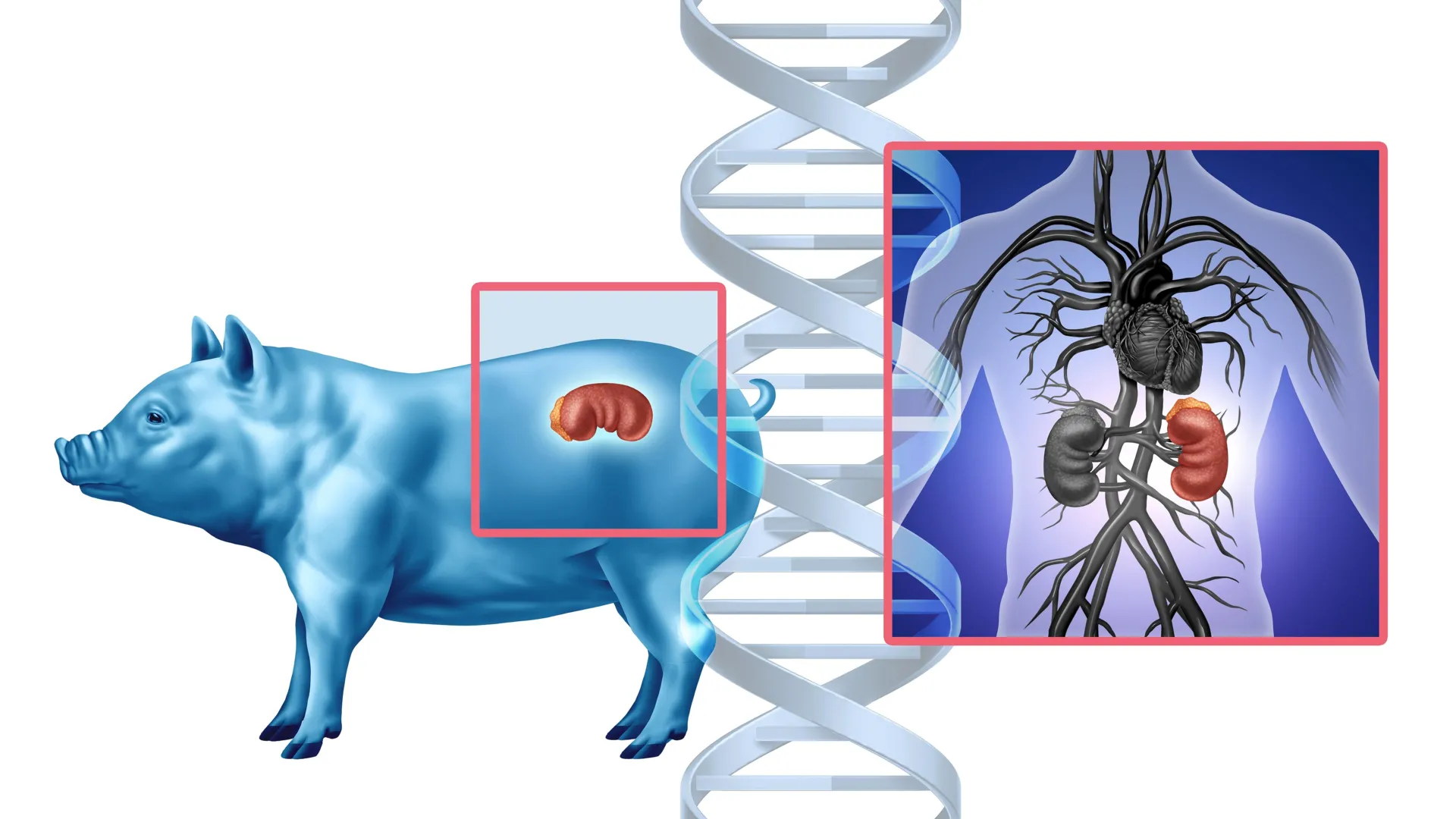
A pioneering study has provided unprecedented insights into the immune response following pig-to-human kidney xenotransplantation.1

The findings, presented today at the ESOT Congress 2025, mark a significant step forward in overcoming the biggest challenge in xenotransplantation: rejection by the human immune system.

Using cutting-edge spatial molecular imaging, researchers mapped how human immune cells interact with pig kidney tissue in transplanted organs, revealing critical early markers of rejection and potential intervention strategies. The study, led by Dr. Valentin Goutaudier and a collaborative international research team (Paris Institute for Transplantation and Organ Regeneration & NYU Langone Transplant Institute), highlights key molecular mechanisms that could shape the future of xenotransplantation.

One of the most striking discoveries was that human immune cells were found in every part of the pig kidney’s filtering system after the transplant. Researchers observed early molecular signs of antibody-mediated rejection as soon as Day 10 and peaking at Day 33, reinforcing previous findings that rejection begins rapidly but progresses over time.2 By tracking these immune responses for up to 61 days, the team identified a crucial window for targeted therapeutic intervention.

“Our study provides the most detailed molecular map to date of how the human immune system engages with a transplanted pig kidney,” explained Dr. Goutaudier. “By pinpointing specific immune cell behaviours and gene expressions, we can refine anti-rejection treatments and improve transplant viability.”

The study’s innovative approach used a bioinformatic pipeline to distinguish human immune cells from pig structural cells, allowing for precise mapping of immune infiltration patterns. Notably, macrophages and myeloid cells were the most prevalent immune cell types across all time points, further confirming their role as key mediators in xenograft rejection.

When targeted therapeutic interventions were introduced, immune-mediated signs of rejection were successfully weakened. Combined with novel spatial insights into how immune cells interact with pig kidney tissue, this marks a major breakthrough — paving the way for more refined anti-rejection strategies. These advances come at a pivotal time as the first US-based clinical trials of pig kidney transplantation into living human recipients begin in 2025.

With xenotransplantation poised to address the global organ shortage crisis, these findings bring researchers one step closer to making genetically modified pig kidneys a viable long-term solution. The next phase will focus on optimising anti-rejection treatments, refining genetic modifications in donor pigs, and developing early detection protocols to monitor and manage rejection responses.

“Understanding the specific immune interactions at a molecular level allows us to develop targeted interventions that can prevent rejection before it escalates,” explained Dr. Goutaudier. “This research lays the groundwork for safer and more effective pig-to-human transplants in the near future.”

As scientific progress accelerates, researchers remain cautiously optimistic that genetically modified pig kidneys could become a routine transplant option within the next decade. However, regulatory approvals will require consistent demonstration of safety and efficacy in diverse patient populations.

References:

  1. Goutaudier V., Williams, C., Morgand, E., et al. Application of a Novel Spatial Transcriptomic 6000-Plex Panel in Pig-to-Human Xenotransplantation. Presented at ESOT Congress 2025; 30th June 2025; London, United Kingdom.
  2. Loupy, A., Goutaudier, V., Giarraputo, A. et al. (2023). Immune response after pig-to-human kidney xenotransplantation: A multimodal phenotyping study.The Lancet, 402(10408), 1158-1169. https://doi.org/10.1016/S0140-6736(23)01855-3
  3. Montgomery RA, Stern JM, Lonze BE, Tatapudi VS, Mangiola M, Wu M, Weldon E, Lawson N, Deterville C, Dieter RA, Sullivan B, Boulton G, Parent B, Piper G, Sommer P, Cawthon S, Duggan E, Ayares D, Dandro A, Fazio-Kroll A, Kokkinaki M, Burdorf L, Lorber M, Boeke JD, Pass H, Keating B, Griesemer A, Ali NM, Mehta SA, Stewart ZA. Results of Two Cases of Pig-to-Human Kidney Xenotransplantation. N Engl J Med. 2022 May 19;386(20):1889-1898. doi: 10.1056/NEJMoa2120238. PMID: 35584156.
Share Button

This virus infects millions—and we just discovered its secret weapon

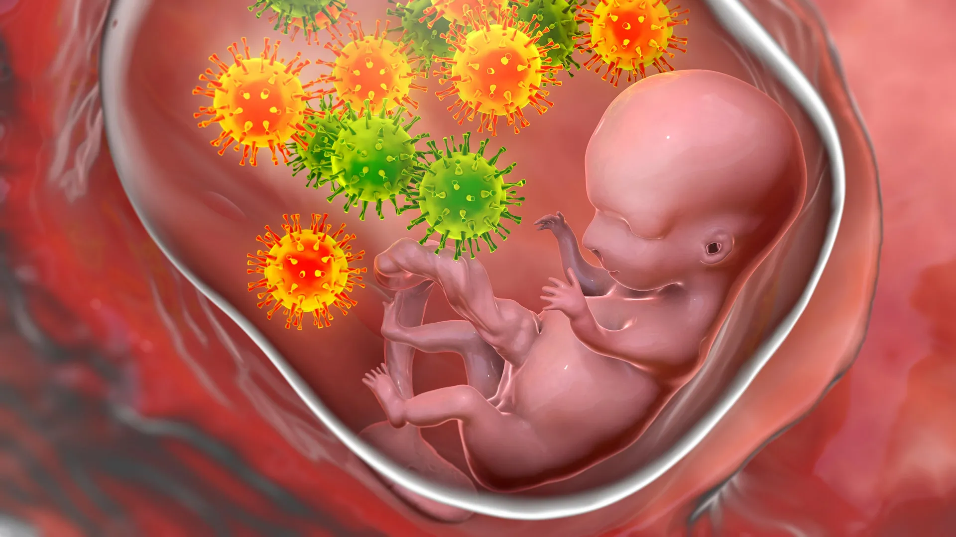
New research from the University of Pittsburgh School of Medicine and La Jolla Institute for Immunology, published today (June 30) in Nature Microbiology, reveals an opportunity for developing a therapy against cytomegalovirus (CMV), the leading infectious cause of birth defects in the United States.

Researchers discovered a previously unappreciated mechanism by which CMV, a herpes virus that infects the majority of the world’s adult population, enters cells that line the blood vessels and contributes to vascular disease. In addition to using molecular machinery that is shared by all herpes viruses, CMV employs another molecular “key” that allows the virus to sneak through a side door and evade the body’s natural immune defenses.

The finding might explain why efforts to develop prophylactic treatments against CMV have, so far, been unsuccessful. This research also highlights a new potential avenue for the development of future antiviral drugs and suggests that other viruses of the herpes family, such as Epstein-Barr and chickenpox, could use similar molecular structures to spread from one infected cell to the next while avoiding immune detection.

“If we don’t know what weapons the enemy is using, it is hard to protect against it,” said senior author Jeremy Kamil, Ph.D., associate professor of microbiology and molecular genetics at Pitt. “We found a missing puzzle piece that represents one possible reason why immunization efforts against CMV have been unsuccessful.”

In the United States, approximately one in every 200 babies is born with congenital CMV infection. Of the babies infected, one in five will have birth defects, such as hearing loss, or go on to have long-term health challenges. For most adults, CMV infections are asymptomatic. But a CMV infection during pregnancy presents significant health risks to the unborn child and could be deadly for people who are immunosuppressed, including organ transplant recipients.

Because of the large size of its genome and its complicated molecular machinery, CMV long evaded attempts to develop prophylactic treatments. Similar to other herpes viruses, CMV relies on a protein called gH to enter cells of the vessel lining. But unlike other herpes viruses, which use a protein partner called gL to facilitate infection, the new study found that CMV replaces gL with another partner called UL116 and recruits a protein called UL141. The resulting complex of gH-UL116-UL141, called GATE by the authors, then becomes an alternative tool for breaking into cells lining the blood vessels and causing internal damage while simultaneously preventing the body’s own immune system from recognizing the signs of infection.

The newly discovered GATE could become a potential vaccine target for CMV and other herpes viruses.

“Previous attempts to generate a CMV vaccine have failed, but that was before we identified the GATE complex. We hope that new strategies targeting GATE will improve our chances to combat CMV infection, and also perhaps cleanse our bodies of this lifelong infection,” said Chris Benedict, Ph.D., associate professor at La Jolla Institute for Immunology and co-senior author of the study with Kamil and LJI professor, president & CEO Erica Ollmann Saphire, Ph.D., MBA. “If we can develop antiviral drugs or vaccines that inhibit CMV entry, this will allow us to combat the many diseases this virus causes in developing babies and immune-compromised people.”

Other authors of this research are Michael Norris, Ph.D., of the University of Toronto; Lauren Henderson, Mohammed Siddiquey, Ph.D., both of Louisiana State University Health Shreveport; and Jieyun Yin, Ph.D., Kwangsun Yoo, Ph.D., Simon Brunel, Ph.D., Michael Mor, Ph.D., and Erica Ollmann Saphire, Ph.D., all of La Jolla Institute for Immunology.

This research was supported by the National Institutes of Health (grants AI11685, AI139749, AI101423 and T32HL155022) and by ARPA-H APECx contract 1AY1AX000055.

Share Button

Parents face hurdles vaccinating children – report

Obstacles include difficulty booking appointments and too few jab reminders, say child health experts.

Share Button

Is RFK Jr’s divisive plan to Make America Healthy Again fearmongering – or revolutionary?

The most powerful public health official in the US is reviled by some medics – but even some of his critics accept that he is bringing drive to healthcare areas that have long been neglected

Share Button

Dads Went On Strike – Now, Parental Leave Might Be Getting An Overhaul

A matter of weeks after the world’s first ‘DadStrike’, where parents gathered to protest the UK’s statutory paternity leave offer, the UK government has announced a review of the parental leave and pay system.

The review will look at the whole system – from maternity and paternity leave to shared parental leave – to see how it can work better for parents and employers.

Views will be gathered from parents, employers and experts across the country, culminating in a roadmap for possible reforms, with the aim being to better support families and grow the economy.

Campaigners have hailed the move as “the best chance in a generation” to improve the system.

Why is it needed?

In a press release, the UK government acknowledged that the current parental leave system is complicated and doesn’t always give families the support they need.

Yet research shows that better parental leave can help close the gender pay gap and boost the economy.

In March this year, mothers opened up about how the UK’s statutory maternity pay offering is not enough to meet the basic costs of living, making it “impossible to live a good life”.

In the UK, statutory maternity pay is paid for up to 39 weeks and parents get 90% of their average weekly earnings (before tax) for the first six weeks, followed by £184.03 or 90% of their average weekly earnings (whichever is lower) for the next 33 weeks.

This is less than half the weekly National Living Wage.

On top of this, analysis of shared parental leave found uptake is skewed towards higher earners – with The Dad Shift, the campaign group behind DadStrike, previously telling the BBC it is “failing working families”.

“Supporting working parents isn’t just the right thing to do – it’s vital for our economy.”

– Angela Rayner

Discussing the new review, deputy prime minister Angela Rayner said: “Those early years are the most special time for families, but too many struggle to balance their work and home lives.

“Supporting working parents isn’t just the right thing to do – it’s vital for our economy.”

The announcement follows a series of changes already progressing in the Employment Rights Bill to improve parental leave and pay.

These include: making paternity leave, unpaid parental leave and flexible working ‘a day one right’; enabling paternity leave and pay to be taken after shared parental leave and pay; and enhancing dismissal protections for pregnant women and new mothers returning to work.

Business secretary Jonathan Reynolds said: “Campaigners have long called for change, and this government has listened. This review is our chance to reset the system and build something that works for modern families and businesses.”

George Gabriel, co-founder of The Dad Shift, told HuffPost UK the review is “the best chance in a generation to improve the system and make sure it actually works for working families”.

“When the last Labour government introduced paternity leave it was groundbreaking. But that offer, unchanged since, is now the least generous in Europe,” he added.

“Our broken parental leave has been overlooked for years, and finally sorting it out would be good not only for parents and children but for businesses too.”

Rachel Grocott, CEO of Pregnant Then Screwed, the campaign group behind 2022′s March of the Mummies, demanding reform of childcare, flexible working and parental leave, said: “It is great to see this long overdue review of the parental leave system. It’s time for the voices of mums, dads, parents and carers everywhere to be heard.

“After 6 weeks mothers are forced to survive maternity leave on 44% less then the National Minimum Wage, and dads are forced to suck up the same benefits for their 2 weeks.

“Yet we know improving parental leave helps children get the best start in life, as well as being better for parents’ heath and equality at home, and closing gender pay and participation gaps in the workplace too.

“Investing in parental leave will pay back above and beyond, to the bottom line and to society: it really is a no-brainer.”

Share Button

More Than 18,000 Russian Troops Reported AWOL Since Putin Invaded Ukraine, UK Says

More than 18,000 Russian soldiers have been reported to the courts for being absent without leave since Vladimir Putin invaded Ukraine, the UK says.

According to the latest update on X from the Ministry of Defence (MoD), 18,159 troops have been faced with a decade-long prison sentence for going AWOL between February 2022 and May 2025.

That is a criminal offence in Russia and a staggering 17,721 (86.2%) of those soldiers were prosecuted.

Citing Russian independent news outlet Mediazona, the MoD said a further 2,379 soldiers were reported to the courts for desertion or refusing orders.

The British officials pointed out there are multiple reasons a soldier could desert.

“Brutal discipline in the Russian Armed Forces, poor medical treatment for injured soldiers and inadequate military training all likely contribute,” the MoD said.

Putin’s soldiers could end up serving on the frontline against Ukraine within 14 days of signing a contract – and just five days of relevant military training.

The MoD continued: “This all runs counter to the official narrative that the Russian leadership values the military service of those who fight in Russia’s illegal war in Ukraine.

“It is a realistic possibility that those prosecuted could commute their sentence by serving in the Russian Storm-Z convict assault units.”

There have been multiple reports of low morale among Russian troops ever since Putin invaded.

In 2023, UK intelligence suggested thousands of Russian casualties in Ukraine were linked to “alcohol consumption” and that soldiers could be “coerced” into joining to boost recruitment numbers.

Despite this chaos within the Russian forces, Putin is still dragging his heels over possible ceasefire talks with Ukraine – even when US mediators suggest terms more favourable to Moscow than Kyiv.

Share Button

Covid inquiry hears of ‘generational slaughter’

Relatives of care home residents tell the Covid inquiry they will never get over how their loved ones died.

Share Button

Scientists just found a sugar switch that protects your brain from Alzheimer’s

A new study from scientists at the Buck Institute for Research on Aging has revealed a surprising player in the battle against Alzheimer’s disease and other forms of dementia: brain sugar metabolism. Published in Nature Metabolism, the research uncovers how breaking down glycogen — a stored form of glucose — in neurons may protect the brain from toxic protein buildup and degeneration.

Glycogen is typically thought of as a reserve energy source stored in the liver and muscles. While small amounts also exist in the brain, particularly in support cells called astrocytes, its role in neurons has long been dismissed as negligible. “This new study challenges that view, and it does so with striking implications,” says Professor Pankaj Kapahi, PhD, senior scientist on the study. “Stored glycogen doesn’t just sit there in the brain; it is involved in pathology.”

The research team, led by postdoc Sudipta Bar, PhD, discovered that in both fly and human models of tauopathy (a group of neurodegenerative diseases including Alzheimer’s), neurons accumulate excessive glycogen. More importantly, this buildup appears to contribute to disease progression. Bar says tau, the infamous protein that clumps into tangles in Alzheimer’s patients, appears to physically bind to glycogen, trapping it and preventing its breakdown.

When glycogen can’t be broken down, the neurons lose an essential mechanism for managing oxidative stress, a key feature in aging and neurodegeneration. By restoring the activity of an enzyme called glycogen phosphorylase (GlyP) — which kicks off the process of glycogen breakdown — the researchers found they could reduce tau-related damage in fruit flies and human stem cell-derived neurons.

Rather than using glycogen as a fuel for energy production, these enzyme-supported neurons rerouted the sugar molecules into the pentose phosphate pathway (PPP) — a critical route for generating NADPH (nicotinamide adenine dinucleotide phosphate) and Glutathione, molecules that protect against oxidative stress. “By increasing GlyP activity, the brain cells could better detoxify harmful reactive oxygen species, thereby reducing damage and even extending the lifespan of tauopathy model flies,” said Bar.

Even more promising, the team demonstrated that dietary restriction (DR) — a well-known intervention to extend lifespan — naturally enhanced GlyP activity and improved tau-related outcomes in flies. They further mimicked these effects pharmacologically using a molecule called 8-Br-cAMP, showing that the benefits of DR might be reproduced through drug-based activation of this sugar-clearing system. “This work could explain why GLP-1 drugs, now widely used for weight loss, show promise against dementia, potentially by mimicking dietary restriction,” said Kapahi.

Researchers also confirmed similar glycogen accumulation and protective effects of GlyP in human neurons derived from patients with frontotemporal dementia (FTD), strengthening the potential for translational therapies. Kapahi says the study emphasizes the power of the fly as a model system in uncovering how metabolic dysregulation impacts neurodegeneration. “Work in this simple animal allowed us to move into human neurons in a much more targeted way,” he said.

Kapahi also acknowledges the Buck’s highly collaborative atmosphere as a major factor in the work. His lab, with expertise in fly aging and neurodegeneration, took advantage of proteomics expertise in the Schilling lab and the Seyfried lab (at Emory University) as well as the Ellerby lab which has expertise in human iPSCs and neurodegeneration.

Kapahi says this study not only highlights glycogen metabolism as an unexpected hero in the brain but also opens up a new direction in the search for treatments against Alzheimer’s and related diseases. “By discovering how neurons manage sugar, we may have unearthed a novel therapeutic strategy: one that targets the cell’s inner chemistry to fight age-related decline,” he says. “As we continue to age as a society, findings like these offer hope that better understanding — and perhaps rebalancing — our brain’s hidden sugar code could unlock powerful tools for combating dementia.”

Coauthors: Additional Buck collaborators include Kenneth A. Wilson, Tyler A.U. Hilsabeck, Sydney Alderfer, Jordan B Burton, Samah Shah, Anja Holtz, Enrique M. Carrera, Jennifer N. Beck, Jackson H Chen, Grant Kauwe, Tara E. Tracy, Birgit Schilling, and Lisa M. Ellerby. Other collaborators include Eric B. Dammer, Fatemeh Seifar and Nicholas T. Seyfried, Emory Center for Neurodegenerative Disease, Emory University School of Medicine, Atlanta, GA as well as Ananth Shantaraman, Department of Biochemistry, Emory University School of Medicine, Atlanta, GA

Acknowledgments: The work was supported by NIH grants R01AG038688, R21AG054121, AG045835, R01AG071995, R01AG070193, T32AG000266-23, R01AG061879, P01AG066591 and 1S10 OD016281. Other support came from the Hevolution Foundation, American Federation of Aging Research, the Larry L. Hillblom Foundation and the CatalystX award from Alex and Bob Griswold

Share Button

This AI tracks lung tumors as you breathe — and it might save lives

In radiation therapy, precision can save lives. Oncologists must carefully map the size and location of a tumor before delivering high-dose radiation to destroy cancer cells while sparing healthy tissue. But this process, called tumor segmentation, is still done manually, takes time, varies between doctors — and can lead to critical tumor areas being overlooked.

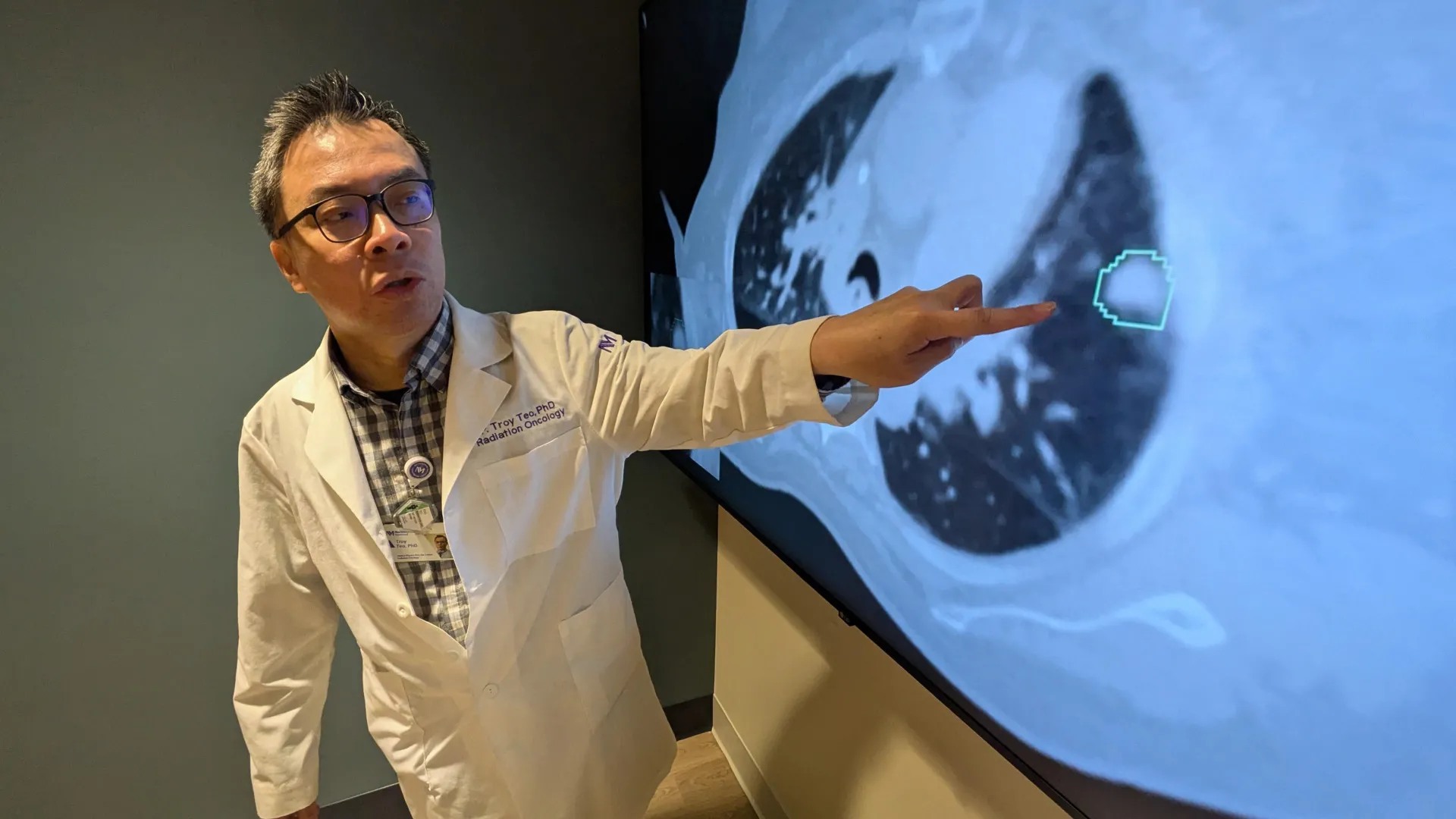
Now, a team of Northwestern Medicine scientists has developed an AI tool called iSeg that not only matches doctors in accurately outlining lung tumors on CT scans but can also identify areas that some doctors may miss, reports a large new study.

Unlike earlier AI tools that focused on static images, iSeg is the first 3D deep learning tool shown to segment tumors as they move with each breath — a critical factor in planning radiation treatment, which half of all cancer patients in the U.S. receive during their illness.

“We’re one step closer to cancer treatments that are even more precise than any of us imagined just a decade ago,” said senior author Dr. Mohamed Abazeed, chair and professor of radiation oncology at Northwestern University Feinberg School of Medicine.

“The goal of this technology is to give our doctors better tools,” added Abazeed, who leads a research team developing data-driven tools to personalize and improve cancer treatment and is a member of the Robert H. Lurie Comprehensive Cancer Center of Northwestern University.

The study was published today (June 30) in the journal npj Precision Oncology.

How iSeg was built and tested

The Northwestern scientists trained iSeg using CT scans and doctor-drawn tumor outlines from hundreds of lung cancer patients treated at nine clinics within the Northwestern Medicine and Cleveland Clinic health systems. That’s far beyond the small, single-hospital datasets used in many past studies.

After training, the AI was tested on patient scans it hadn’t seen before. Its tumor outlines were then compared to those drawn by physicians. The study found that iSeg consistently matched expert outlines across hospitals and scan types. It also flagged additional areas that some doctors missed — and those missed areas were linked to worse outcomes if left untreated. This suggests iSeg may help catch high-risk regions that often go unnoticed.

“Accurate tumor targeting is the foundation of safe and effective radiation therapy, where even small errors in targeting can impact tumor control or cause unnecessary toxicity,” Abazeed said.

“By automating and standardizing tumor contouring, our AI tool can help reduce delays, ensure fairness across hospitals and potentially identify areas that doctors might miss — ultimately improving patient care and clinical outcomes,” added first author Sagnik Sarkar, a senior research technologist at Feinberg who holds a Master of Science in artificial intelligence from Northwestern.

Clinical deployment possible ‘within a couple years’

The research team is now testing iSeg in clinical settings, comparing its performance to physicians in real time. They are also integrating features like user feedback and working to expand the technology to other tumor types, such as liver, brain and prostate cancers. The team also plans to adapt iSeg to other imaging methods, including MRI and PET scans.

“We envision this as a foundational tool that could standardize and enhance how tumors are targeted in radiation oncology, especially in settings where access to subspecialty expertise is limited,” said co- author Troy Teo, instructor of radiation oncology at Feinberg.

“This technology can help support more consistent care across institutions, and we believe clinical deployment could be possible within a couple of years,” Teo added.

This study is titled “Deep learning for automated, motion- resolved tumor segmentation in radiotherapy.”

Share Button

Ancient DNA reveals leprosy hit the Americas long before colonization

  • Hansen’s Disease (leprosy) is thought to have originated in Eurasia: previous studies on Mycobacterium leprae, the dominant form of leprosy, suggest the disease originated in Eurasia.
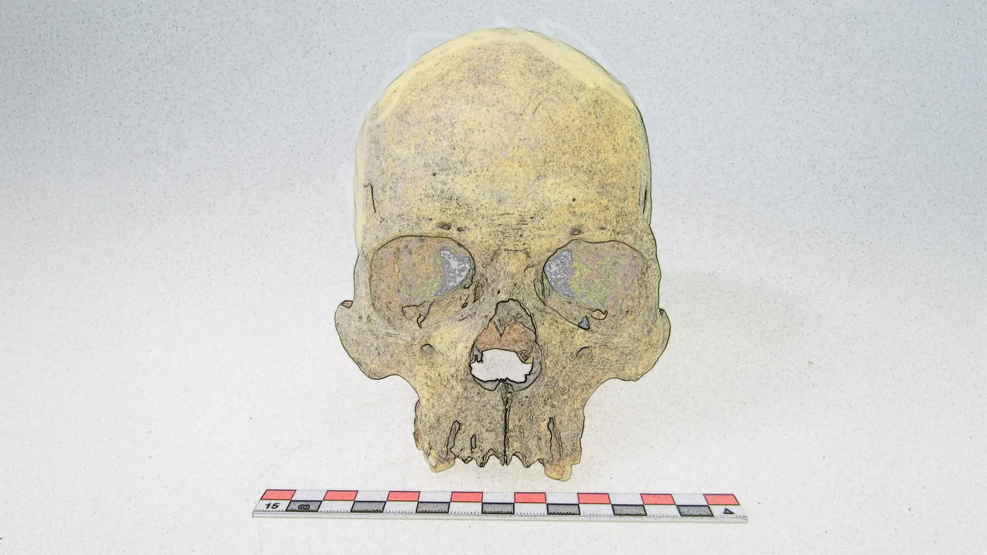
  • Ancient pathogen genomes from old bones: a team of scientists from Germany and Argentina have reconstructed two genomes of Mycobacterium lepromatosis in 4000-year-old human skeletons from Chile. This pathogen is regarded as a second, less common, cause of Hansen’s Disease.
  • A new American chapter for Hansen’s Disease: two pathogens that are responsible for the same disease evolved separately on opposite sides of the world for thousands of years.

Hansen’s Disease, more commonly known as leprosy, is a chronic disease that can lead to physical impairment. Today it exists in over 100 countries, and while the infection is treatable, access to treatment varies widely with socioeconomic conditions. Its mention in historical texts give us a glimpse into its past impact on population health in Europe and Asia. Prolonged untreated infection can result in characteristic changes in bone, and these have been documented in archaeological skeletons as early as 5000 years ago in Europe, Asia, and Oceania. So far, absence of these characteristic changes in the pre-contact American contexts suggests that leprosy was one of the many diseases introduced to the continent in the colonial period. Thereafter it afflicted humans and curiously also armadillos.

From a genetic perspective, Hansen’s Disease is caused by either the globally dominant Mycobacterium leprae or the newly identified and rare Mycobacterium lepromatosis. The recovery of M. leprae from archaeological bone in Europe suggests the disease originated in Eurasia sometime during the Neolithic transition about 7000 years ago. Similar emergence estimates have been proposed for other notorious diseases such as plague, tuberculosis, and typhoid fever. Ancient genomes for M. lepromatosis have remained elusive, and these may hold important clues on the history of Hansen’s Disease.

Past disease in the American continent

We know comparatively little about the infectious disease experience of the diverse communities of people living in the Americas before the colonial period. This accounts for almost 20,000 years of human history, and the diverse ecosystems into which humans integrated across the continent would have presented challenges to the immune system not otherwise encountered in other parts of the world. We know very little of these diseases, as they were smothered by the onslaught of pathogens that Europeans later brought with them. Archaeological study of human bones from the pre-contact Americas confirm that the time was far from disease-free, but often the traces seen on the bones aren’t specific enough to assign to a known disease.

“Ancient DNA has become a great tool that allows us to dig deeper into diseases that have had a long history in the Americas,” says Kirsten Bos, group leader for Molecular Paleopathology at the Max Planck Institute for Evolutionary Anthropology. She and her team have been studying pathological bone from American context for over a decade. Some diseases that the group has found were expected – just last year they found evidence that the family of diseases closely related to syphilis had its roots in the Americas, which many had suspected. “The advanced techniques now used to study ancient pathogen DNA allows us to look beyond the suspects and into other diseases that might not be expected from the context,” she adds.

Re-writing the history of Hansen’s Disease

Bos’s team worked closely with researchers from Argentina and Chile to both identify bones suitable for analysis and to carry out the meticulous work of isolating the DNA of ancient pathogens. Doctoral candidate Darío Ramirez of the University of Córdoba, Argentina, worked extensively with such material, and was the first to identify a genetic signature related to leprosy in some 4000-year-old skeletons from Chile. “We were initially suspicious, since leprosy is regarded a colonial-era disease, but more careful evaluation of the DNA revealed the pathogen to be of the lepromatosis form.” This provided the first clue that M. lepromatosis and M. leprae, though nominally both pathogens that cause Hansen’s Disease, might have very different histories. Reconstruction of the genome was key in looking into this. While putting the molecular puzzle of an ancient genome back together is never an easy task, these pathogens in particular had “amazing preservation, which is uncommon in ancient DNA, especially from specimens of that age,” comments Lesley Sitter, a postdoctoral researcher in Bos’s team who carried out the analysis.

The pathogen is related to all known modern forms of M. lepromatosis, but as there are so few genomes available for comparison, there is still much to be learned about it. This work has shown that a pathogen considered rare in a modern context caused disease for thousands of years in the Americas. Rodrigo Nores, professor of Anthropology at the University of Córdoba, Argentina is convinced that more cases, both ancient and modern, will be identified in the coming years: “this disease was present in Chile as early as 4000 years ago, and now that we know it was there, we can specifically look for it in other contexts.” Once more genomes surface, we’ll be able to piece out further details of its history and better understand its global distribution today. The pathogen has recently been discovered in squirrel populations from the United Kingdom and Ireland, but in the Americas it has yet to be found in any species other than humans. With such little data, mystery still surrounds its origin. “It remains to be determined if the disease originated in the Americas, or if it joined some of the first settlers from Eurasia,” adds Bos. “So far the evidence points in the direction of an American origin, but we’ll need more genomes from other time periods and contexts to be sure.”

Share Button