Physicists discover a hidden gluon structure inside protons that could rewrite textbooks

New findings from the STAR detector at the Relativistic Heavy Ion Collider (RHIC) are challenging a familiar picture of what gives protons one of their defining quantum properties. The results suggest that gluons, the particles that act as the glue holding quarks together, may play a key role in carrying and conserving baryon number.

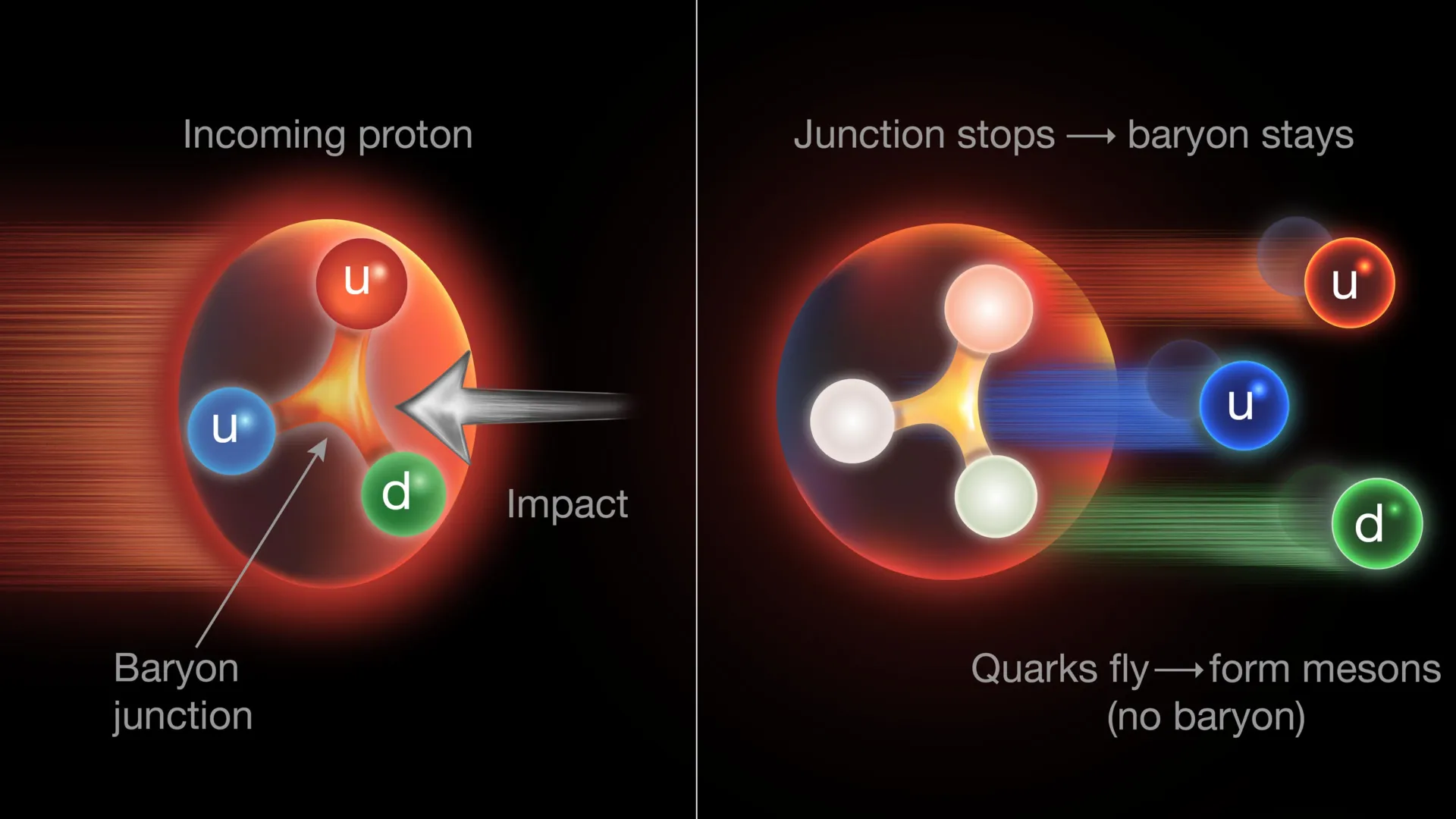
The evidence comes from high-energy particle collisions at RHIC, a U.S. Department of Energy (DOE) Office of Science user facility for nuclear physics research that operated at DOE’s Brookhaven National Laboratory from 2000 to early 2026. According to the new study, published in Science, baryon number may be associated with a Y-shaped “junction” of gluons connecting the proton’s three main quarks. If confirmed, that would challenge the long-standing assumption that baryon number belongs exclusively to those quarks.

“Traditionally, scientists have assumed that each of the three main ‘valence’ quarks inside a proton or neutron carries one-third of the baryon number,” said Zhangbu Xu, a professor at Kent State University with a joint appointment at Brookhaven Lab.

A Decades-Old Idea About Gluons

Physicists first proposed the baryon junction, also called a gluon junction, in the 1970s as a way to describe how gluons connect the valence quarks inside a proton. In 1996, four years before RHIC began operating, Dmitri Kharzeev, a theoretical physicist at Stony Brook University and Brookhaven Lab, proposed that this junction might do something even more fundamental. Rather than the valence quarks carrying baryon number, the junction itself could be responsible.

The STAR collaboration has now developed a way to test that possibility using several types of collisions produced at RHIC.

“Using data collected from different types of particle collisions at RHIC, our results suggest that the baryon number is not simply carried by individual quarks,” Xu added. “Our findings strongly support the idea that baryon number is more favorably carried and transported by gluons, the particles that hold quarks together, when arranged in this special configuration.”

Why Baryon Number Matters

Determining what actually carries baryon number matters far beyond the internal structure of a proton. In RHIC collisions, conservation of baryon number means that the total number of baryons, three-quark particles such as protons and neutrons, must remain unchanged before and after the collision. The same conservation principle also applies on the scale of the universe.

“Since the Big Bang, the number of protons and neutrons all together never changes as a function of time,” said Nicole Lewis, a STAR physicist at Rice University who started this project as a postdoc at Brookhaven Lab in 2020. “The reasons for this conservation are not well understood. It’s one of the mysteries of the universe, related to why we have more matter than antimatter,” she said.

Baryon number conservation also has a much more tangible consequence. It helps explain the extraordinary stability of protons, which form a central part of atomic nuclei and do not appear to decay under ordinary circumstances.

“It’s believed that the lifetime of a proton is longer than the lifespan of the universe,” Lewis said. “This allows atomic nuclei to form and be stable — which means matter, as we interact with it in the universe, can exist.”

A More Complicated Proton

The possibility that gluons carry baryon number would overturn the standard simplified description found in many textbooks. In that picture, a proton has a baryon number of plus one, divided equally among its three main valence quarks. Each quark therefore carries plus one third of the baryon number, much as the proton’s electric charge is distributed among its three valence quarks.

But real protons are much more complicated than that simplified model suggests.

“In the naïve quark model, there are three quarks inside a proton, but nothing else,” said Tommy Tsang, formerly a postdoc at Kent State University, now at DOE’s Argonne National Laboratory. “But if we look at details inside, there are not only three quarks but also a lot of gluons interacting, connecting between those quarks, and there are also quarks and antiquarks that pop up from the vacuum, so it’s actually a really complex object.”

Quantum chromodynamics (QCD), the theory used to describe these interactions, has been highly successful in explaining the strong force that acts among quarks and gluons. Even so, models inspired by QCD often need additional assumptions to reproduce some of the particle patterns observed when RHIC smashes nuclei together at nearly the speed of light.

An Unexpected Excess of Baryons

One observation in particular caught the STAR team’s attention. The detector repeatedly records more baryons than antibaryons emerging sideways from the collisions, perpendicular to the direction of the incoming beams.

“In the STAR detector, we consistently see an excess of baryons coming out of the collisions perpendicular to the direction of the colliding beams,” Tsang said. “The fact that we end up with more baryons than antibaryons — or more matter than antimatter — is not surprising since our collisions start with matter,” he said.

These extremely energetic collisions convert tremendous amounts of energy into thousands of newly created particles. What puzzled the researchers was not simply that more baryons than antibaryons were produced. It was where the excess baryons appeared.

If valence quarks alone carried the baryon number, explaining the excess away from the beamline would require all three valence quarks from one colliding proton to stop near the center of the detector. They would then have to undergo a conversion from matter into energy and back into matter, producing new baryons that move outward perpendicular to the beam.

The STAR researchers suspected there might be another explanation.

Electric Charge Provides a Test

The team found a way to investigate the mystery by taking advantage of another property of valence quarks: electric charge. Scientists compared the net baryon number measured in different RHIC nuclear collisions with the way electric charge was redistributed in those same events.

“Measuring the electric charge coming out perpendicular to the collision gives you a definitive way of measuring how many quarks are stopped and transformed into new particles,” said Zebo Tang, a professor at the University of Science and Technology of China who led a group of students performing data analyses and model simulations.

The comparison revealed a striking mismatch. Researchers observed roughly twice as many baryons as should have been produced based on the electric charge associated with stopped quarks.

According to models based on QCD, that means too few quarks were being stopped to account for all the baryons appearing in the detector.

That left an important question: What was carrying the extra baryon number?

The STAR physicists argue that gluons offer a possible answer, specifically the three-pronged gluon junction that connects the proton’s valence quarks.

How the Gluon Junction Could Carry Baryon Number

The proposed mechanism depends on what happens when protons inside colliding nuclei reach enormous energies. According to the STAR team, the “gluon junction” or “baryon junction” that links the quarks may be much easier to stop in a collision than the three quarks themselves.

If the junction is stopped, its energy can be converted into newly produced baryons that travel outward in directions perpendicular to the beams. Meanwhile, the valence quarks that were previously connected by the junction can continue moving forward along the beampipe.

Understanding why requires looking at the changing internal structure of a proton as its energy increases.

“The baryon junction is always there even as protons are accelerated to higher and higher energy,” Prithwish Tribedy, a STAR physicist at Brookhaven Lab. “But at high energy, gluons within the proton split and multiply.”

As the number of gluons increases, the proton’s momentum becomes spread among more of them. Each individual gluon, including those forming the junction, therefore carries a smaller portion of the proton’s total momentum. The valence quarks, however, continue to carry much of the proton’s forward motion.

As a result, when the collision occurs, the comparatively slower three-pronged gluon junction should be easier to stop and convert into new particles than the rapidly moving quarks.

Stopping one connected structure is also simpler than stopping three separate quarks, making such an interaction more likely, according to Tribedy.

“In the collision, the baryon junction gets held behind, and the quarks continue on,” he noted.

Building New Particles After the Collision

Quarks and gluons cannot remain isolated, so after the collision they quickly combine with other particles.

In a simplified example, a quark continuing down the beampipe could join with an antiquark and form a two-quark particle called a meson. At the same time, the three-pronged gluon junction could behave somewhat like a Y-shaped magnet, drawing in three newly created quarks from the vacuum and producing a new baryon.

Actual RHIC collisions are considerably more violent and complex.

“Even though we start with nuclei that contain roughly 100 protons and 100 neutrons, these collisions create thousands of new particles; 99% of the energy is transformed into new particles,” said Rongrong Ma, a Brookhaven Lab physicist.

The STAR team found that collisions producing larger numbers of particles also showed a greater excess of “midrapidity” baryons compared with predictions based on the simpler picture in which quarks alone carry baryon number.

The fact that so many of these baryons emerge perpendicular to the beamline provides strong evidence, according to the researchers, that the baryon junction exists and plays an important role in transporting baryon number.

Rethinking a Fundamental Property of Matter

The results suggest that one of the proton’s defining quantum properties may not reside solely in its three valence quarks. Instead, the gluon structure connecting those quarks could be central to how baryon number is carried through energetic collisions.

“Our research challenges the long-held idea that baryon number is simply divided among and carried by the three quarks,” said Ma. “This new understanding reshapes how we think about the structure of matter and deepens our knowledge of the most fundamental element that is responsible for the universe in its current form.”

The research was supported by the DOE Office of Science, the U.S. National Science Foundation (NSF), and numerous international agencies and organizations listed in the scientific paper. Researchers also used the Open Science Grid, which is supported directly by NSF, along with computing resources at Brookhaven Lab’s Scientific Data and Computing Facilities and the National Energy Research Scientific Computing Center (NERSC), another DOE Office of Science user facility located at DOE’s Lawrence Berkeley National Laboratory.

Share Button

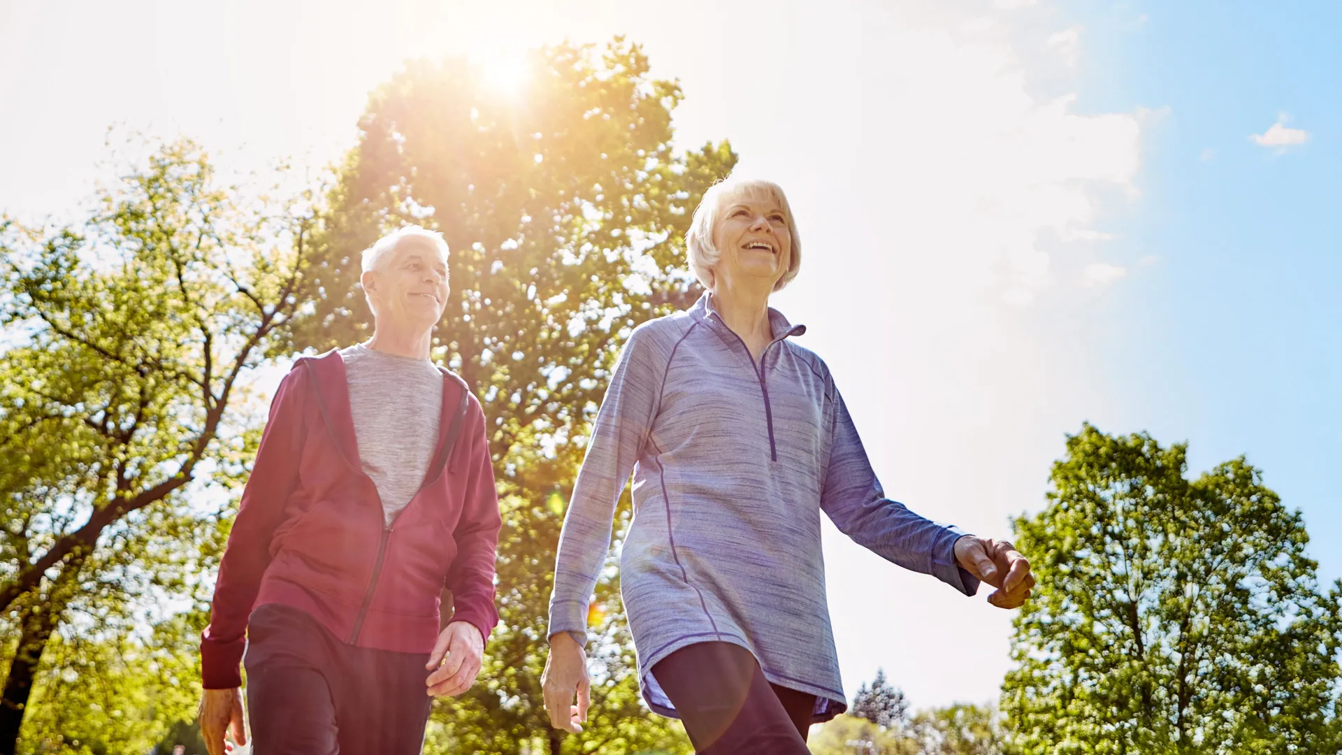
Scientists reveal why walking gets so much harder with age

New Australian research is shedding light on why walking often becomes slower and more tiring with age. The findings suggest that the body gradually gives up some movement efficiency in favor of staying stable and upright.

Led by researchers at Flinders University and the University of Canberra, the study found that aging is associated with a “safety-first” walking strategy. This approach emphasizes stability instead of speed and energy efficiency, helping explain why older adults may fatigue more quickly and also face a greater risk of falling.

Researchers analyzed movement data from 107 healthy adults between the ages of 26 and 86. They found small but meaningful age-related differences in how the ankle and nearby muscles manage each step.

How the Ankle Changes With Age

Lead author and sport and exercise technology expert Dr. Cody Lindsay says the ankle is essential for maintaining balance while also helping propel the body forward.

“As we get older, the body starts to favor stability over efficiency,” says Dr. Lindsay, from the Flinders Caring Futures Institute.

“That helps keep us upright, but it also makes walking more of an effort.”

The researchers found that older adults are more likely to activate opposing muscles around the ankle at the same time. This pattern is known as co-contraction. It makes the ankle joint stiffer and can improve stability when the foot contacts the ground.

However, Dr. Lindsay says this comes at a cost.

“Stiffening the joint makes walking safer, but it also means the muscles are working harder without generating as much forward movement,” he says.

Older participants also generated less push-off power with each step. As a result, their strides were shorter, and their walking speeds were slower.

A Safety First Strategy for Walking

Co-author Associate Professor Maarten Immink says the findings point to a wider change in the way the body manages movement as people grow older.

“The nervous system adopts a safety-first approach, compensating for age-related changes by favoring stability over performance,” says Associate Professor Immink, Lead of the Active Lives Research Program within the Caring Futures Institute at Flinders University.

“These changes can also increase fatigue and make walking longer distances more challenging, while reducing the ability to recover from trips or slips — a key factor in falls among older adults.”

“Even gradual changes can affect confidence and independence, and people may notice they tire more quickly or feel less steady, especially on uneven ground.”

Exercise May Help Preserve Mobility

The findings also suggest possible ways to help people maintain mobility as they age.

Rather than focusing only on building strength, the researchers say exercise programs should also emphasize balance and coordination while paying attention to how different muscles work together during each step.

“For older Australians, simple actions can make a difference, including regular physical activity, balance exercises such as tai chi, lower-leg strengthening and activities that challenge coordination,” says Dr. Lindsay, from Flinders’ College of Health and Enablement.

“Staying active is one of the most important things people can do, and small, consistent exercises can help you stay confident, mobile and independent for longer.”

The researchers hope these findings can contribute to improved prevention and rehabilitation strategies designed to reduce falls and support healthy aging.

Share Button

I Added A Baking Staple To My Chickpeas, And It Made Them Silkier And Creamier Than Ever

I’ve been banging on about the merits of slow cookers, especially during a heatwave, all summer.

Even though they’re usually associated with stodgy winter dishes, their lower temperature and enhanced insulation mean they don’t warm your kitchen up as much as you’d expect.

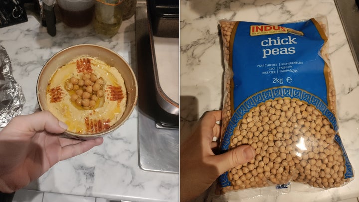
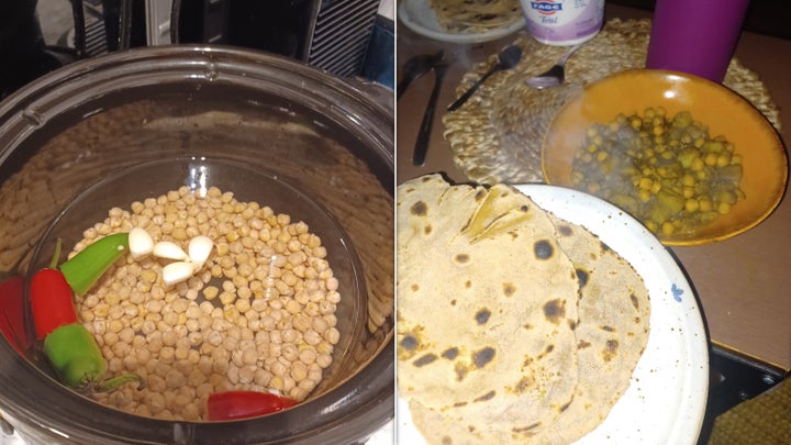
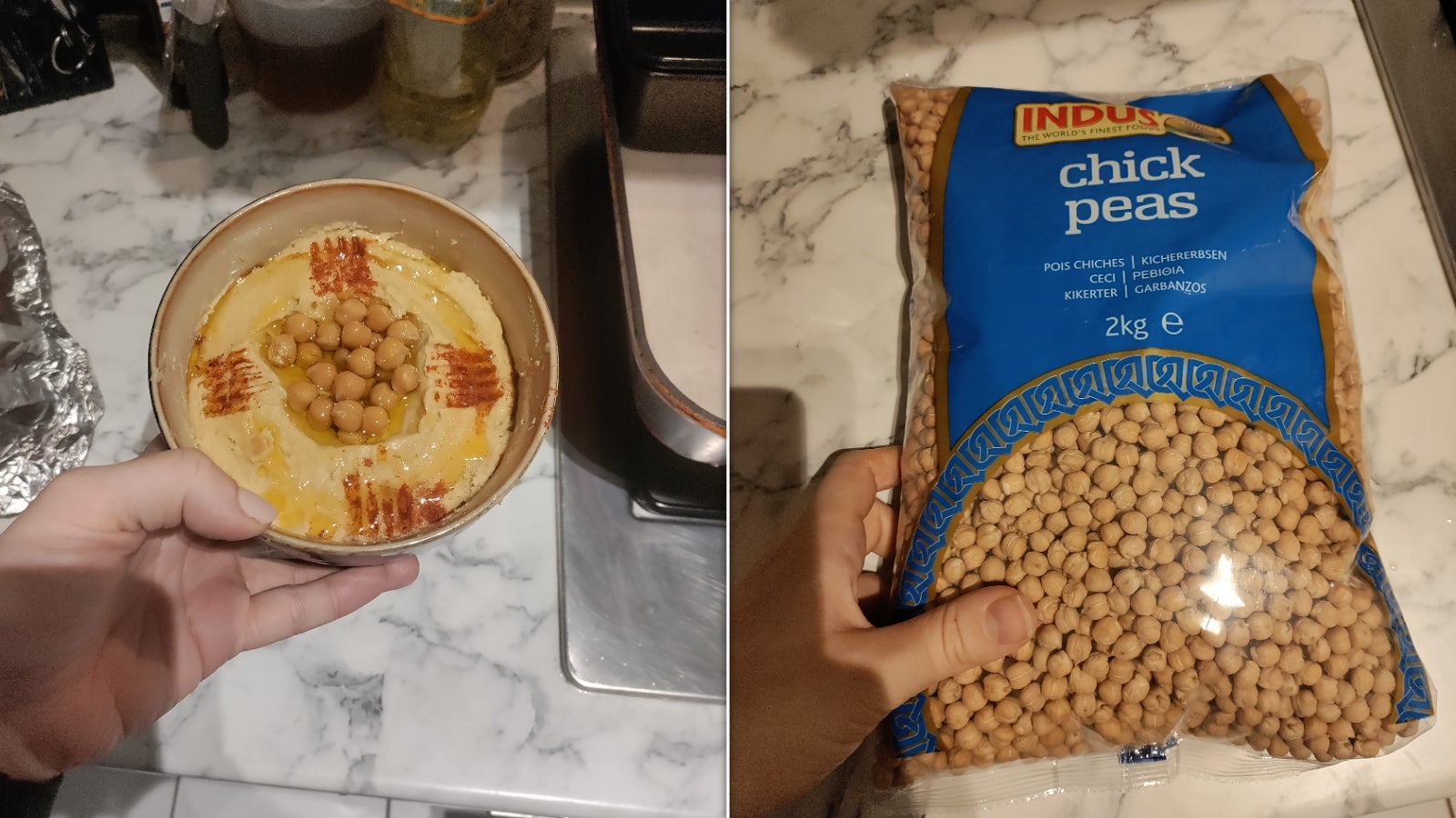
Alongside a delicious coconut lentil soup and some Greek-inspired mains, I’ve been batch-cooking my own chickpeas, too. (Yes, canned is easier, but tinned chickpeas are also designed to keep their shape in the container, meaning they’re never quite soft enough for dishes like hummus).

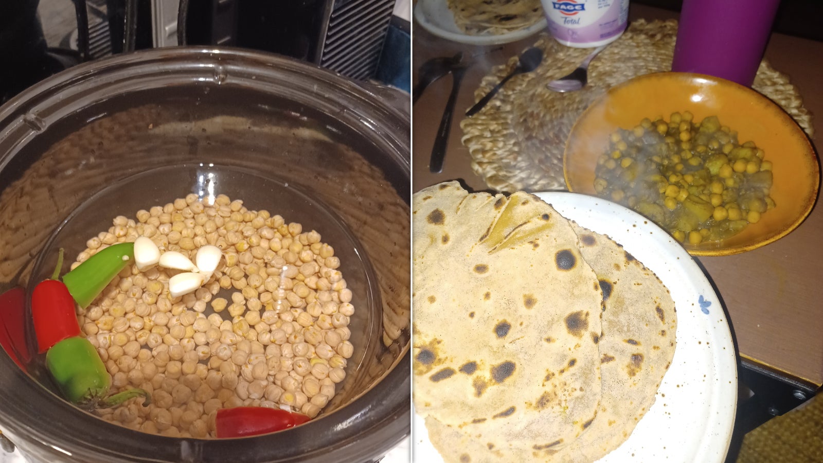
Still, it wasn’t until last week that I tried a long-standing trick to make them softer, creamier, and more delicious – and I wish I’d started years before.

Add baking soda to your chickpeas

Hummus, chickpeas

Amy Glover / HuffPost UK

Hummus, chickpeas

I usually add garlic, salt, and chilli to my chickpea water for a bit of added flavour. Salt might have a small advantage in softening them, but for the best results, you need baking soda.

There’s some interesting science behind the addition. When you add the baking staple to chickpea water, it slowly replaces the legumes’ rigid calcium ions with bendier sodium ions.

And the alkaline nature of baking soda also breaks down the bonds that hold their pectins (a type of plant fibre) together.

Combined, those factors mean that baking soda makes chickpeas soft, squidgy, and almost fudgy.

Their skins also soften and even begin to flake away depending on how long you cook them. That, I find, is especially useful for hummus – though I like a looser, softer chickpea for things like salads and chickpea curries, too.

Chickpeas in the slow cooker: then in a curry

Amy Glover / HuffPost UK

Chickpeas in the slow cooker: then in a curry

I was amazed by how well the trick worked. Compared to chickpeas I’d stewed for hours, those cooked with baking soda became softer much faster – and eliminated that harder centre I could never quite coax into silky perfection.

How much baking soda should I add to my chickpeas?

You don’t need much to see impressive results. I use about a teaspoon per 500g of dry chickpeas, but some recipes halve that.

The amount you’ll need to add depends, partly, on how long you’re cooking chickpeas for. If you’re making them on the hob, more baking soda will be better; however in the slow cooker, where they stew for hours, try less.

Serious Eats notes that you don’t have to spend hours on the method if you don’t have time. You can soften tinned chickpeas using baking soda, too: just drain them, add enough liquid to cover them, and whack in some baking soda while they boil. Half a teaspoon per 400g can of chickpeas should do the job.

Test the chickpeas as they cook to make sure they haven’t collapsed completely. Once they’ve reached the desired texture, you can remove them and drain them.

This has an added benefit: the chickpea water from baking-soda-cooked legumes is a bit bitter and unpleasant. So, if you need to use this for your recipe, you can save the liquid from the can and use that later on.

Share Button

Why Gen Z women know more about periods than their mums

From PMOS to cycle syncing, young women are teaching their mums about periods.

Share Button

What’s driving men to get penis fillers, despite the risks?

Men who have had the procedure can expect 10-20% more girth for the next six to nine months – but it can go wrong.

Share Button

Scientists detect a surprising shift in human blood as CO2 rises

Rising levels of carbon dioxide in the atmosphere may already be influencing human biology. New research has identified long-term changes in blood chemistry that appear to track rising atmospheric CO2, raising concerns that an important blood marker could approach the upper end of its healthy range within the next several decades.

The findings may be particularly important for children and teenagers. Because their bodies are still developing, younger generations are expected to experience the greatest lifetime exposure to elevated atmospheric CO2.

Decades of Blood Data Reveal a Shift

In a study published in Air Quality, Atmosphere and Health, scientists from The Kids Research Institute Australia, Curtin University and The Australian National University (ANU) examined more than 20 years of U.S. population health data. They found persistent changes in several measures of blood chemistry that closely followed the upward trend in atmospheric CO2.

The researchers used information from the U.S. National Health and Nutrition Examination Survey (NHANES), analyzing blood test results from roughly 7,000 people at two-year intervals between 1999 and 2020.

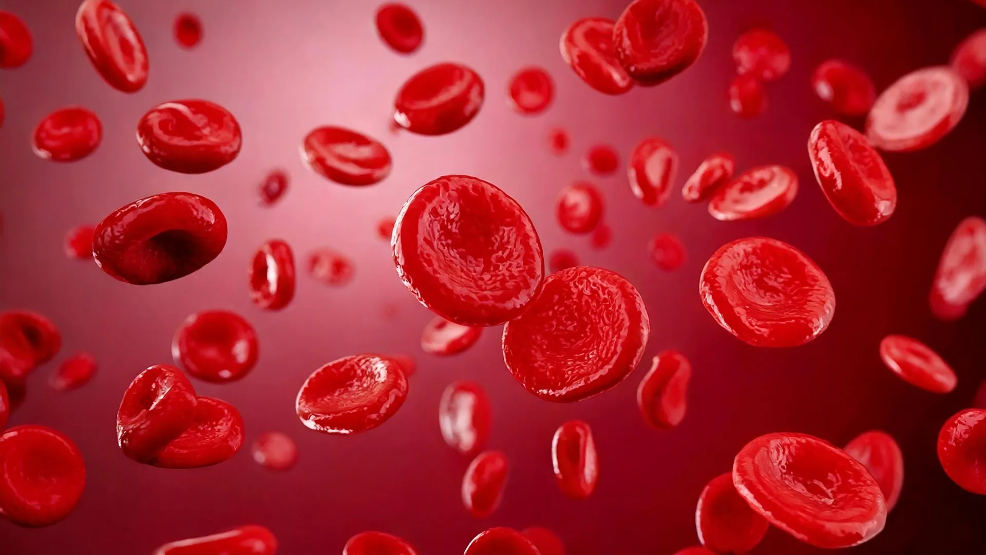
Since 1999, average serum bicarbonate levels have increased by about 7 percent. Bicarbonate is a blood marker closely associated with carbon dioxide in the body. During the same period, average levels of calcium and phosphorus decreased.

Those biological trends occurred as atmospheric CO2 climbed from about 369 parts per million (ppm) in 2000 to more than 420 ppm today.

Study author Associate Professor Alexander Larcombe said the results indicate that the body may already be adjusting to changes in the composition of the atmosphere.

“What we’re seeing is a gradual shift in blood chemistry that mirrors the rise in atmospheric carbon dioxide, which is driving climate change,” A/Prof Larcombe said.

How the Body Responds to More CO2

Bicarbonate is essential for regulating the body’s acid-base balance. As CO2 increases, the body can retain additional bicarbonate to help keep blood pH stable. Although this response helps preserve that balance, maintaining it over long periods could have physiological effects.

“If current trends continue, modeling indicates average bicarbonate levels could approach the upper limit of today’s accepted healthy range within 50 years,” A/Prof Larcombe said

“Calcium and phosphorus levels could also reach the lower end of their healthy ranges later this century.”

Humans evolved when atmospheric CO2 concentrations were approximately 280 to 300 ppm. During the past decade, atmospheric levels have risen by an average of about 2.6 ppm each year, while 2024 alone saw an increase of 3.5 ppm.

Fellow Author Dr Phil Bierwirth, a retired environmental geoscientist affiliated with the ANU Emeritus Faculty, emphasized that the study does not establish a direct cause-and-effect relationship. However, he said the consistency of the changes across a large population warrants attention.

“I actually think that what we are seeing is because our bodies are not adapting,” Dr Bierwirth said.

“It appears we are adapted to a range of CO2 in the air that may now have been surpassed.

“The normal range maintains a delicate balance between how much CO2 is in the air, our blood pH, our breathing rate and bicarbonate levels in the blood.

“As CO2 in the air is now higher than humans have ever experienced, it appears to be building up in our bodies. Maybe we can never adapt such that it is vitally important to limit atmospheric levels of CO2.”

A Potential New Dimension of Climate Risk

The researchers say the results suggest that rising atmospheric CO2 could represent a form of climate-related risk that is different from more familiar threats such as heatwaves, extreme weather and sea-level rise.

According to A/Prof Larcombe, increasing CO2 may need to be considered not only as an environmental concern, but also as a long-term public health factor that should be monitored.

“We’re not saying people are suddenly going to become unwell when we cross a certain threshold,” he said.

“But this suggests there may be gradual physiological changes occurring at a population level, and that’s something we should be monitoring as part of future climate change policy.”

The researchers recommend monitoring the composition of the atmosphere together with biological markers across populations. Tracking both alongside established climate indicators could help scientists determine how slow environmental changes affect human biology over periods of decades.

CO2 Reduction Could Have Health Implications

Cutting CO2 emissions remains essential for limiting global warming. The findings also raise the possibility that lowering emissions could have an additional role in protecting long-term human health.

The researchers argue that potential physiological effects from rising CO2 should therefore be considered in future discussions about climate policy, alongside its established environmental consequences.

Associate Professor Larcombe is part of the Wal-yan Respiratory Research Centre, a partnership between The Kids Research Institute Australia, Perth Children’s Hospital and Perth Children’s Hospital Foundation.

Share Button

When gut microbes run low on fiber, they may start eating you

Plant based diets support health in several ways, including benefits for the gut, immune system, metabolism, and cardiovascular system. Part of that effect comes from encouraging a diverse population of bacteria in the intestines. Scientists have long known that dietary fiber contributes to these benefits because gut microbes help break it down. Plants also contain colorful “phytochemicals” that help protect them from environmental threats and may influence human health. Even so, researchers are still working to understand exactly how gut bacteria process the many components of plant foods and how those interactions produce beneficial effects.

Two studies led by Ludwig Princeton’s Jenna AbuSalim and Director Joshua Rabinowitz offer new insight into that process. One appears in the current issue of the Proceedings of the National Academy of Sciences, while the other was published in Nature Metabolism in June. The first study found that plant fiber and certain plant proteins can change microbial metabolism in ways that increase beneficial metabolites while reducing harmful ones. The second showed that several biologically important metabolites usually credited to gut microbes can also be produced in substantial amounts by mammalian metabolism.

“There’s growing interest across medical disciplines in manipulating the human microbiome or using its metabolic products themselves for therapy,” said Rabinowitz. “Diet holds great promise for controlling the microbiome and its outputs. But to devise effective therapeutic interventions, we need to understand what aspects of the diet control which microbial outputs.”

How Plant Foods Shift Gut Metabolites

In the PNAS study, Rabinowitz, AbuSalim and their colleagues examined how plant based foods influence phenol metabolites. Gut bacteria create these compounds when they digest the amino acids tyrosine and phenylalanine, but the resulting metabolites can have very different effects on health.

Phenylpropionate and hippuric acid are produced when bacteria process phenylalanine, and they are associated with gut health and healthy body weight. By contrast, p-cresol sulfate and phenol sulfate come from tyrosine and have been linked to worse outcomes in cancer patients as well as systemic toxicity in people with kidney disease.

“Our studies showed that both the fiber and indigestible proteins from plants — which we call ‘proteins imitating fiber,’ or Prif — shift the balance of phenol metabolites from the harmful kind made from tyrosine to the healthful variety derived from phenylalanine,” said AbuSalim.

Fiber has long been recognized as an important part of a healthy diet, but indigestible plant proteins have received far less attention. AbuSalim, Rabinowitz and their colleagues found that these proteins are processed by gut microbes and can alter both the makeup of the microbiome and the host’s metabolism. Working together with indigestible plant fiber, they can also change the metabolic activity of gut bacteria in ways that favor the production of beneficial phenols.

When Gut Bacteria Turn to the Gut Lining

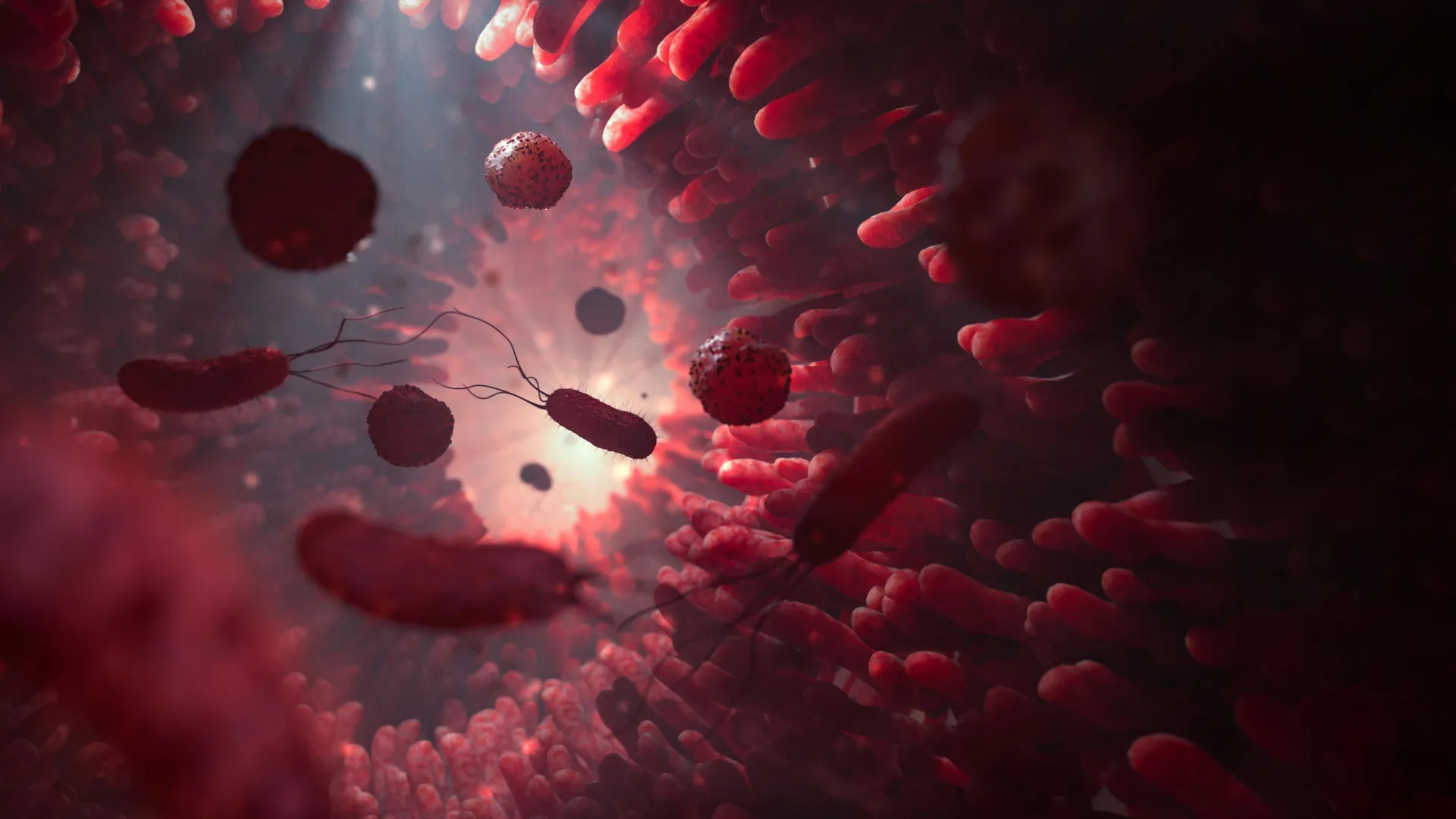
To trace where these compounds came from, the researchers labeled proteins with stable (non-radioactive) isotopes and followed their digestion in the mouse gut. They found that the “bad” phenols were produced when bacteria consumed proteins from the host, including proteins found in the mucus lining of the gut. The good phenols, in contrast, came almost entirely from indigestible proteins in the diet (Prif).

Fiber reduced the bacterial breakdown of the gut’s mucus lining, which in turn lowered production of the harmful phenols. Prif increased the amount of dietary protein that reached gut microbes, giving them more material to produce the beneficial phenols.

“We think Prifs represent an emerging class of dietary nutrients that shape the composition of the gut microbiome and could have a far-reaching influence on metabolic health,” said AbuSalim.

“Food packaging may eventually list Prif right below fiber,” said Rabinowitz.

Rethinking Where Gut Metabolites Come From

The Nature Metabolism study focused on the origins of phenol metabolites as well as indole metabolites, which are produced from the amino acid tryptophan. Like phenols, indoles are being studied for their possible therapeutic value.

Indole metabolites have been connected to a wide range of diseases, including inflammatory bowel disease, neurodegenerative disorders, and cancer. In cancer research, they have been found to affect processes that include cancer metastasis and anti-tumor immune responses.

Scientists had generally assumed that phenols and indoles were produced only by gut bacteria. AbuSalim, Rabinowitz and their colleagues decided to test that assumption. Researchers have been especially interested in dietary and probiotic approaches that might increase beneficial indole metabolites. But those strategies may need to be reconsidered if mammalian metabolism, rather than microbes, is responsible for much of what circulates in the body.

Using isotope tracing in mice, rats and human cells, the researchers found that mammalian metabolism can produce many indole and phenol metabolites on its own. These included important compounds such as indole-3-lactate and indole-3-acetate.

In mice, circulating levels of these metabolites remained high even after antibiotic treatment disrupted the microbiome. A similar pattern appeared in samples from patients taking antibiotics, including cancer patients. At the same time, metabolites made exclusively by microbes, including indole-3-propionate and p-cresol sulfate, declined after antibiotic treatment.

New Clues for Diet and Microbiome Therapies

Together, the two studies provide a clearer picture of where phenol and indole metabolites come from and how they are produced. The findings could influence the development of therapies designed to raise or lower specific metabolites.

They also add important detail to scientists’ understanding of how diet interacts with the microbiome. Knowing which foods influence particular microbial products could eventually help researchers design more precise dietary, probiotic, or metabolic interventions.

“Beyond that,” said Rabinowitz, “a clearer picture of how different foods interact with the microbiome to modulate the production of bacterial metabolites will help sharpen the guidance nutritionists and doctors can give to people for disease prevention and therapy.”

These studies were funded by the Ludwig Institute for Cancer Research, the National Institutes of Health, the National Institute of Diabetes and Digestive and Kidney Diseases, the Princeton Alliance for Collaborative Research and Innovation, Princeton University.

Aside from his post as Director of the Princeton Branch of the Ludwig Institute for Cancer Research, Joshua Rabinowitz is Professor in the Department of Chemistry & Lewis-Sigler Institute for Integrative Genomics and a member of the Rutgers Cancer Institute.

Share Button

Scientists solve the mystery of a brain “switch” that can trigger weight loss in opposite ways

Cambridge researchers have uncovered why both activating and blocking the same brain receptor can promote weight loss. The findings may help scientists develop obesity treatments that are more effective and potentially work better in combination.

The mouse study, published in Nature Metabolism, found that the outcome depends on which part of the brain is targeted. Activating the receptor in the brainstem reduced appetite, while blocking the same receptor in the hypothalamus produced a similar weight loss effect through a different mechanism.

More than a billion people around the world are living with obesity, a condition that raises the risk of diseases including type 2 diabetes, cardiovascular disease and cancer. Losing weight can reduce some of these risks, but achieving substantial weight loss through diet and exercise alone can be difficult.

How Modern Weight Loss Drugs Target the Brain

A new generation of weight loss medications has emerged in recent years that act on specific receptors involved in appetite. By influencing these receptors, the drugs can reduce food intake, promote weight loss, and help regulate blood sugar.

Several widely used medications, including Wegovy and Ozempic, activate a protein receptor called the glucagon-like peptide 1 receptor (GLP-1R).

Other obesity treatments act on both GLP-1R and another receptor known as the glucose-dependent insulinotropic polypeptide receptor (GIPR). This second target has presented scientists with an unusual puzzle.

Some medications, including Mounjaro and Zepbound, activate GIPR. Others, such as MariTide, block it. Despite producing opposite effects on the same receptor, both approaches can help promote weight loss.

Researchers at the Institute of Metabolic Science, University of Cambridge, set out to understand why. Their experiments in mice revealed that the two types of GIPR drugs work through different regions of the brain. The researchers also found that these approaches can increase weight loss when paired with certain GLP-1-based weight loss medicines.

Tracking GIPR Activity in Different Brain Regions

To identify the brain regions responsible for these effects, the team used genetically engineered mice in which GIPR had been selectively removed from specific areas.

One group lacked GIPR in the brainstem, the region at the base of the brain just above the spinal cord that is involved in appetite and nausea. Another group lacked the receptor in the hypothalamus, an important brain region involved in regulating hunger and body weight. A third group consisted of normal, unmodified mice that served as controls.

The scientists treated the animals with different combinations of a GIPR agonist (which activates the receptor), a GIPR antagonist (which blocks the receptor) and a GLP-1 drug. They then monitored food consumption, body weight, fat mass, blood sugar control and brain activity.

Comparing the different groups allowed the team to pinpoint where each treatment was acting.

The results showed that GIPR agonists primarily work through the brainstem. Activating GIPR in this region reduced appetite and led to lower body weight.

Blocking a Brain Brake on Fullness

GIPR antagonists followed a different route.

Instead of acting primarily through the brainstem, the researchers found that blocking GIPR promoted weight loss through the hypothalamus. In this region, GIPR appears to function as a kind of ‘brake’ that limits how strongly the brainstem responds to signals indicating that the body is full.

Blocking the receptor effectively releases that ‘brake’, allowing fullness signals to have a stronger effect.

The researchers also found evidence that blocking GIPR could enhance the effects of emerging medicines that target the amylin receptor. This suggests that GIPR antagonists might eventually be useful for strengthening several different classes of obesity treatments.

Clues to More Powerful Obesity Drug Combinations

The results help explain why treatments such as MariTide can be effective. MariTide, currently in phase 3 clinical trials, combines GIPR antagonism with GLP-1 receptor agonism.

Understanding how these separate pathways interact could also help researchers design more effective combinations of obesity medicines in the future.

Dr. Jo Lewis, the study’s first author from the Institute of Metabolic Science at the University of Cambridge, said: “Understanding which brain circuits respond to these medications – and how they do so – could help us design better drugs that produce more weight loss with fewer side effects, and which might work in combination with other obesity medicines to even greater effect.

“Our work also strengthens the idea that the brain is central to obesity treatment. Obesity drugs are not acting simply on the gut or pancreas. Instead, they have important effects on specific, identifiable brain circuits that regulate appetite and food intake.”

The research was funded by the Medical Research Council and Wellcome.

Share Button

The Common Travel Sleep Mistake Giving You A ‘Holiday Sleep Hangover’

For some people, the joy of a holiday is slightly dampened by the inevitable impacts holidaying can have on sleep. In fact, according to new research commissioned by sleep technology firm Simba has found that over three quarters of Brits 76% find that holiday travel affects their sleep, with over 2 in 5 UK adults finding that tiredness affects the first day of their holiday and more.

Very relatable. The first day of every holiday for me seems to be experienced in a tired and wired daze.

It doesn’t stop there, though. In fact, this tiredness can reoccur once you’re back home in what sleep experts have dubbed a “Holiday Sleep Hangover”.

The ‘Holiday Sleep Hangover’

According to Lisa Artis, Deputy CEO of Simba’s charity partner, The Sleep Charity, the “holiday sleep hangover” describes the lingering effects of travel-related sleep disruption caused by early airport starts and poor in-flight sleep to unfamiliar hotel rooms, changing time zones and disrupted routines

But unlike a traditional hangover, it can strike twice: “once when you arrive on holiday and again when you return home.”

“The holiday sleep hangover has become the hidden physical and mental cost of modern travel,” says Artis.

“Sleep disruption usually starts much earlier and can continue long after you’ve unpacked your suitcase. It’s the combination of lost sleep, changes to routine and, for some travellers, jet lag that can leave people feeling out of sync for days.”

Additionally, if you’re a sucker for an early flight so you can “make the most of the day” once you arrive at your destination, you may be doing yourself a disservice, Artis warns, saying: “If you’re up at 3am for an airport transfer after a restless night’s sleep, you’re already asking your body to function on less recovery than it normally would. That sleep debt can follow you into the first few days of your trip.”

“It can be tempting to over-optimise the night before you travel. But trying to force a really early night can make it harder to switch off,” she explains.

“You end up clock-watching and feeling more alert.”

Instead, she recommends keeping your usual evening routine calm and consistent.

How to recover from a Holiday Sleep Hangover

According to Lisa, the most effective approach is getting back to a consistent routine as quickly as possible. That means returning to regular bedtimes, exposing yourself to daylight during the day and avoiding the temptation to stay up late to prolong the holiday feeling.

“Your sleep environment can help speed up recovery. When you’re trying to get your routine back on track, comfort and temperature become important,” adds Arts.

“If you’re waking because you’re too hot, too cold or uncomfortable, it can make recovery feel slower.”

Take care of yourself!

Share Button

It Was Too Late To Fix My Relationship With My Dying Father, So I Taught Children To Read Instead

I hadn’t seen my dad in five years when my sister texted me that he had fallen at home. After I had my first child, the childhood abuse I had previously overlooked to keep the peace suddenly became incomprehensible, and I chose to become estranged from him.

But when I found out he was dying, I knew time was running out to reconcile. I didn’t want to regret missing it.

On my first visit to see him, I brought flowers and kneeled by his bedside. He was thin, jaundiced and bedridden. His fine hair was snowy and delicate over his scalp, his movie star cheekbones more prominent than ever. His brown eyes still looked like mine.

But he didn’t recognise me. He stared at me and looked away repeatedly, his expression bewildered and hurt.

“Who are you?” he asked.

I was stunned and told him who I was.

“Is it OK that I’m here, or would you prefer I go?” I asked.

“You can stay. I’m just surprised. It’s been a long time.” There was grief and accusation in his voice.

“I know,” I said.

I wouldn’t apologise for the estrangement. He wouldn’t apologise for the abuse. We wouldn’t talk about the past when death was so close.

When I left, I kissed his forehead and promised I’d be back.

A few weeks later, I signed up to be an early literacy volunteer, teaching young children to read. Once a week, I spent an hour with the students during their lunch break, reviewing the alphabet and helping them sound out words while they ate orange slices and whole wheat pizza.

Then I would spend an hour with my dad at the healthcare facility where he lived for his last five months. We would share a few minutes of small talk, I would feed him small bites of baked fish or tater tots, and we’d watch TV.

The two things were connected. My father’s side of the family is full of educators. My dad was a professor for more than 50 years. He’s the reason I have post-traumatic stress disorder, but he’s also the reason I love literature, travel, art, food and languages. His mother – my namesake – was once a missionary-teacher who rode on horseback four miles every day to reach her students.

“Teaching is a spiritual thing,” my dad said.

I thought volunteering to help children read would connect me to my dad and our family as he was dying. The kids were at the beginning of their lives and education, while my father, the professor, was coming to the end of his.

I volunteered on Mondays. Usually, I visited my father at the end of the week. My weeks were measured in the bookends of life.

There were six schools where I could volunteer – all underfunded and with low reading test scores. I chose the one closest to my childhood home, my dad’s house. I was a kindergartener in that neighbourhood the first time my dad squeezed my wrists so hard that bruises encircled them for weeks.

The school was a rectangular building made with dark bricks. I had passed by it countless times as a child. From the outside, it looked institutional in the late September sun. But inside, I was surprised by how light it was. The hallways were painted white and worn in places with scuffs and pencil squiggles – imprints from students growing up.

Three large trees grew in the foyer outside the library. Their shiny green leaves wove a canopy over the grey and blue tiles where kids walked from recess, lunch and library time. That’s where I met the kids with whom I’d be working, affectionately called Book Buddies.

The volunteer program coordinator handed me two folders, each bearing the name of a little boy. We had two assignments for the day. The first was to complete “getting to know me” sheets. The second was to assess how many letter names and sounds they knew.

I asked them their birthdays, their favorite colour and to draw their favourite food. My first buddy knew the letters in his name but no others. My second buddy surprised me by knowing all the letters and their sounds, but he said them quietly with his eyes downcast.

The author volunteering for the Book Buddies program

Photo Courtesy Of Virgie Townsend

The author volunteering for the Book Buddies program

At the school, there was the squeak of sneakers as kids ran in the hall and the voices of teachers announcing it was time to line up. There were boxes of small books, word games with dice, pencils, plastic utensils and thin straws for chocolate milk cartons.

Over time, I learned my first buddy loved to take a big sip of chocolate milk and burp and then we’d laugh together. He introduced me to his kindergarten friends. In the winter, his boot shoelaces were always undone, so I tied them before he returned to class.

My second buddy rarely made eye contact but read everything I put in front of him perfectly. He was reserved, but told me he lived with his dad and loved reading books the program gave him.

I praised both kids for their reading, hard work and the progress they made.

Sometimes after volunteering, I’d drive by my childhood home. With my father gone, the house stood empty.

At the healthcare facility, there was a crane to lift my dad, who used to hike by the Sequoias near his own childhood home. There were nurses who joked as they spooned medication into his mouth and the blare of televisions playing at full volume. There were cartons of Ensure and automatic hand sanitiser dispensers.

Sometimes my dad seemed stronger. On a good day, we watched Star Trek, and he laughed out loud for the first time in a while. When I left, he called after me in a strong voice, “Love you, honey.”

But organ failure is a rollercoaster with many ups and downs, all heading toward the inescapable end. Mostly, I worried about the emergence of new symptoms like the jaundice and itchiness that came and went. The bones I hadn’t seen before, jutting clavicle and breastbone. Lost fat on his fingers, every knuckle visible beneath the thin skin.

On a cold January day, I was with my dad when he began seizing. The seizures were quick, full-body and terrifying. He didn’t remember them – he just thought he’d fallen asleep for a minute. The doctor said there was nothing else they could do.

Four days later, he was unconscious. His breaths were long, slow and gasping. His TV was off. He had stopped responding to us. I asked my sisters for a minute alone with him. Everything we had left was in that small room.

“I hope that in whatever comes next, you receive the love you didn’t get as a child and you’re able to give that love to yourself and others in return,” I told him. “If there’s regret or anything else you’re carrying, you can put it by the wayside. You don’t have to carry it for me.”

He died the next morning. After he took his last breath, I watched the pulse in his neck grow fainter until I couldn’t see it beating under his skin anymore.

“Dad, I’m just going to check your pulse, OK?” I said. I placed two fingers on his neck – a place I had never touched before – and felt no movement.

Before the funeral home director placed him on the gurney, I kissed his forehead one final time and then wept.

Depleted by grief and the paperwork that accompanies a parent’s death, I didn’t return to volunteering for a month. Sometimes I’d look at a photo of my dad from when he was 5 years old – the same age as my own son and my volunteering buddies.

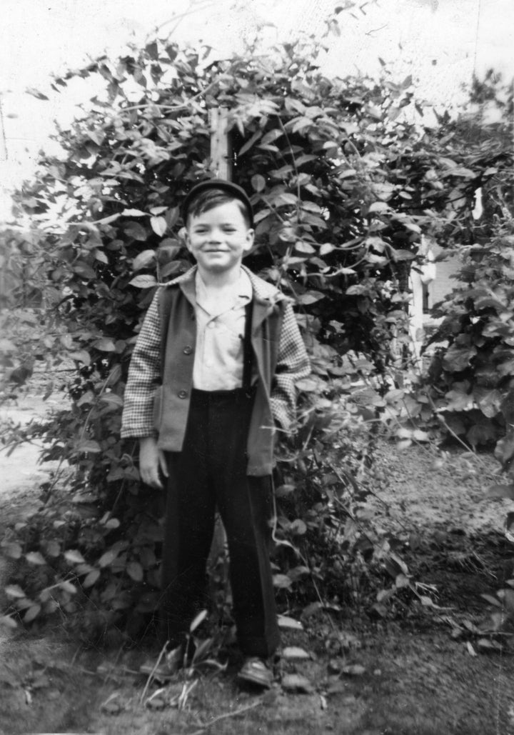
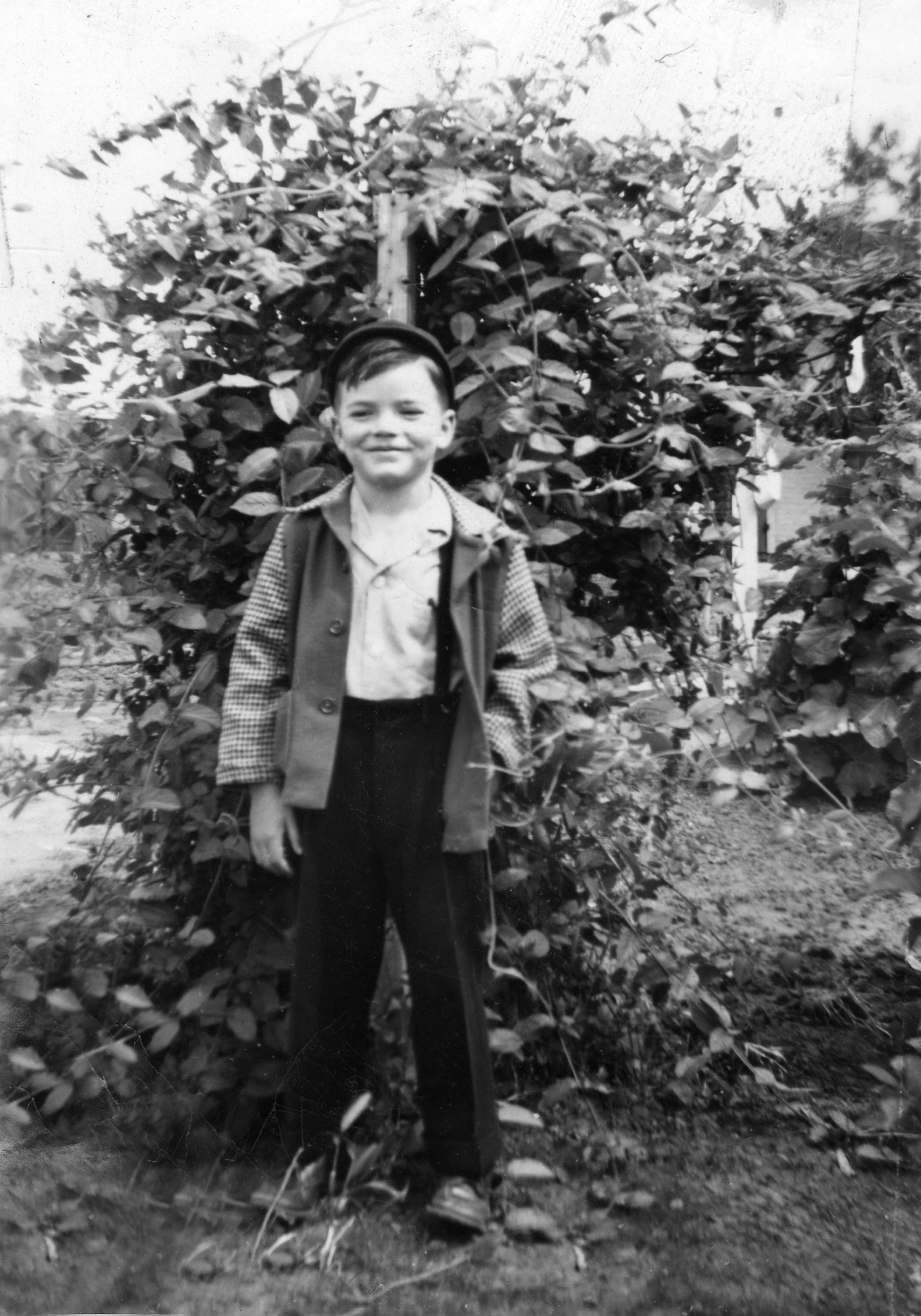
In the picture, my dad wore a black cap over his dark hair, and his left hand was in the pocket of his checkered coat. Although I knew he had an unsafe, loveless childhood, he beamed a proud smile. I wished I could go back in time to protect that little boy.

The author's father, John Townsend, at 5 years old

Photo Courtesy Of Virgie Townsend

The author’s father, John Townsend, at 5 years old

When I went back to the school, my first buddy walked up to me with his lunch tray and a mildly scolding expression on his face.

“Where have you been?” he asked, as if I were a child who missed curfew.

“One of my family members was sick,” I said, smiling. “I’m back now.”

I was glad to be in the school, reading with the kids under the trees in the foyer. One day, as I arrived, my buddy was standing in the lunch line. He saw me, exclaimed to a friend, “That’s my Book Buddy!” and ran to hug me.

Later, when my second buddy caught a cold, I gave him tissues to take back to class because I knew he’d be too shy to ask the teacher. I helped another student carefully cut out a booklet for herself to take home to read. I made sure one of their classmates didn’t leave behind her lunchbox.

Little acts of care I wish had been given to the children that my father and I once were.

On the last day of volunteering, we held a party in the school library. I helped my buddies don plastic gold medals and paper graduation hats. We gave them backpacks with books, a toy keychain, bubbles and a red bracelet that said, “I love to read.”

“Do you love to read?” I asked my buddy, who was now reading several levels ahead of where he began.

He looked down bashfully and nodded with a smile, his two front teeth missing. We listened to a story about joyriding farm animals and played sight word bingo with candy. Then I tied his shoelace one last time, wished him a great summer, and watched him walk back to class.

I realised that while a child cannot heal their own parent, I could give that care to my kids and to other children who needed it. It was too late for my dad, but it might be right on time for them.

Do you have a compelling personal story you’d like to see published on HuffPost? Find out what we’re looking for here and send us a pitch at pitch@huffpost.com.

Help and support:

If you, or someone you know, is in immediate danger, call 999 and ask for the police. If you are not in immediate danger, you can contact:

  • The Freephone 24 hour National Domestic Abuse Helpline, run by Refuge: 0808 2000 247
  • In Scotland, contact Scotland’s 24 hour Domestic Abuse and Forced Marriage Helpline: 0800 027 1234
  • In Northern Ireland, contact the 24 hour Domestic & Sexual Violence Helpline: 0808 802 1414
  • In Wales, contact the 24 hour Life Fear Free Helpline on 0808 80 10 800.
  • National LGBT+ Domestic Abuse Helpline: 0800 999 5428
  • Men’s Advice Line: 0808 801 0327
  • Respect helpline (for anyone worried about their own behaviour): 0808 802 4040
Share Button