When our vertical perception gets distorted: Body pitch and translational body motion

Our ability to perceive what is truly vertical is crucial. Without it, we would struggle to perform simple tasks such as holding a cup of coffee without spilling it and maintaining appropriate body posture.

Now, an international team of scientists from Japan, Canada and Germany have discovered a new situation from our everyday environment where this ability is compromised — when our body pitches (body pitch) and moves at the same time (body motion).

The scientists’ observations, which were published in the journal Multisensory Research, were based on real-world scenarios, revealing factors long overlooked by past laboratory studies.

“Unlike typical studies of this kind, which happen in well-controlled laboratories, our team traveled to Hong Kong to conduct field research on the world-renowned Peak Tram,” said Dr Chia-huei Tseng, who led the research and is associate professor at Tohoku University’s Research Institute of Electrical Communication (RIEC). “We performed experiments on hundreds of commuters making their way up Hong Kong’s highest hill, Victoria Peak.”

Although body pitch and body motion have been studied in previous studies, the phenomena were isolated. This study combined both aspects to determine a new situation where verticality becomes distorted.

The team enlisted the help of an architect to build a device that participants could hold to indicate their perceived verticality (i.e., subjective haptic vertical or SHV).

In most laboratory setting, humans are capable of perceiving SHV with an error of less than 1 degree. However, when measurements were taken on the moving Peak Tram, a misjudgment of 10 degrees was observed, something never previously reported.

To investigate the possible sources of errors, the researchers devised additional experiments. First, they had participants close their eyes to rule out visual influences. Then, to eliminate the effects of the vestibular system — the sensory system that creates a sense of balance and spatial orientation — they inserted a back wedge between the participants and their chairs. None of these alternations had much bearing on diminishing the SHV error, ruling out visual and vestibular systems as the primary cause.

Additionally, they ran their tests on Hong Kong’s Ding Ding Tram, which runs at similar speeds to the Peak Tram but on a relatively flat surface. In both situations, no bias was detected.

Psychology Professor Kenzo Sakurai from Tohoku Gakuin University, an expert on self-motion and co-author of the study, points out what a major discovery their findings are. “The failure to accurately sense our body orientation relative to gravity may result in inappropriate movements or falls, as well as impair daily functions such as walking, climbing a staircase, and carrying a tray. In specialized tasks such as driving a car or piloting a plane, this failure could be disastrous.”

Dr Tseng thinks that their study demonstrates a great example of scientific inquiry generalizing the actual world we live in. “On top of its pure scientific contribution, this study challenges the misconception that research is limited to scientific cubicles indoors. In this case, the natural environment provided a well-designed puzzle for scientists to solve.”

With international travel restriction gradually lifting, the paper’s authors encourage those to take a ride on Hong Kong’s Peak Tram to feel this fascinating perceptual phenomenon. But maybe wait until after the ride to get your coffee.

Story Source:

Materials provided by Tohoku University. Note: Content may be edited for style and length.

Share Button

Good and bad feelings for brain stem serotonin

New insights into the opposing actions of serotonin-producing nerve fibres in mice could lead to drugs for treating addictions and major depression.

Scientists in Japan have identified a nerve pathway involved in the processing of rewarding and distressing stimuli and situations in mice.

The new pathway, originating in a bundle of brain stem nerve fibres called the median raphe nucleus, acts in opposition to a previously identified reward/aversion pathway that originates in the nearby dorsal raphe nucleus. The findings, published by scientists at Hokkaido University and Kyoto University with their colleagues in the journal Nature Communications, could have implications for developing drug treatments for various mental disorders, including addictions and major depression.

Previous studies had already revealed that activating serotonin-producing nerve fibres from the dorsal raphe nucleus in the brain stem of mice leads to the pleasurable feeling associated with reward. However, selective serotonin reuptake inhibitors (SSRIs), antidepressant drugs that increase serotonin levels in the brain, fail to exert clear feelings of reward and to treat the loss of ability to feel pleasure associated with depression. This suggests that there are other serotonin-producing nerve pathways in the brain associated with the feelings of reward and aversion.

To further study the reward and aversion nerve pathways of the brain, Hokkaido University neuropharmacologist Yu Ohmura and Kyoto University pharmacologist Kazuki Nagayasu, together with colleagues at several universities in Japan, focused their attention on the median raphe nucleus. This region has not received as much research attention as its brain stem neighbour, the dorsal raphe nucleus, even though it also is a source of serotonergic nerve fibres.

The scientists conducted a wide variety of tests to measure activity of serotonin neurons in mice, in response to stimulating and inhibiting the median raphe, by using fluorescent proteins that detect entry of calcium ions, a proxy of neuronal activation in a cell-type specific manner.

They found that, for example, pinching a mouse’s tail — an unpleasant stimulus — increased calcium-dependent fluorescence in the serotonin neurons of the median raphe. Giving mice a treat such as sugar, on the other hand, reduced median raphe serotonin fluorescence. Also, directly stimulating or inhibiting the median raphe nucleus, using a genetic technique involving light, led to aversive or reward-seeking behaviours, such as avoiding or wanting to stay in a chamber — depending on the type of stimulus applied.

The team also conducted tests to discover where the switched-on serotonergic nerve fibres of the median raphe were sending signals to and found an important connection with the brain stem’s interpenduncular nucleus. They also identified serotonin receptors within this nucleus that were involved in the aversive properties associated with median raphe serotonergic activity.

Further research is needed to fully elucidate this pathway and others related to rewarding and aversive feelings and behaviours. “These new insights could lead to a better understanding of the biological basis of mental disorders where aberrant processing of rewards and aversive information occur, such as in drug addiction and major depressive disorder,” says Ohmura.

Story Source:

Materials provided by Hokkaido University. Note: Content may be edited for style and length.

Share Button

Enzyme that protects against viruses could fuel cancer evolution

An enzyme that defends human cells against viruses can help drive cancer evolution towards greater malignancy by causing myriad mutations in cancer cells, according to a study led by investigators at Weill Cornell Medicine. The finding suggests that the enzyme may be a potential target for future cancer treatments.

In the new study, published Dec. 8 in Cancer Research, scientists used a preclinical model of bladder cancer to investigate the role of the enzyme called APOBEC3G in promoting the disease and found that it significantly increased the number of mutations in tumor cells, boosting the genetic diversity of bladder tumors and hastening mortality.

“Our findings suggest that APOBEC3G is a big contributor to bladder cancer evolution and should be considered as a target for future treatment strategies,” said study senior author Dr. Bishoy M. Faltas, assistant professor of cell and developmental biology at Weill Cornell Medicine, and an oncologist who specializes in urothelial cancers at NewYork-Presbyterian/Weill Cornell Medical Center.

The APOBEC3 family of enzymes is capable of mutating RNA or DNA — by chemically modifying a cytosine nucleotide (letter “C” in the genetic code). This can result in an erroneous nucleotide at that position. The normal roles of these enzymes, including APOBEC3G, are to fight retroviruses like HIV — they attempt to hobble viral replication by mutating the cytosines in the viral genome.

The inherent hazardousness of these enzymes suggests that mechanisms must be in place to prevent them from harming cellular DNA. However, starting about a decade ago, researchers using new DNA-sequencing techniques began to find extensive APOBEC3-type mutations in cellular DNA in the context of cancer. In a 2016 study of human bladder tumor samples, Dr. Faltas, who is also director of bladder cancer research at the Englander Institute for Precision Medicine and a member of the Sandra and Edward Meyer Cancer Center, found that a high proportion of the mutations in these tumors were APOBEC3-related — and that these mutations appeared to have a role in helping tumors evade the effects of chemotherapy.

Such findings point to the possibility that cancers generally harness APOBEC3s to mutate their genomes. This could help them not only acquire all the mutations needed for cancerous growth but also boost their ability to diversify and “evolve” thereafter — enabling further growth and spread despite immune defenses, drug treatments, and other adverse factors.

In the new study, Dr. Faltas and his team, including first author Dr. Weisi Liu, a postdoctoral research associate, addressed the specific role of APOBEC3G in bladder cancer with direct cause-and-effect experiments.

APOBEC3G is a human enzyme not found in mice, so the team knocked out the gene for the sole APOBEC3-type enzyme in mice, replacing it with the gene for human APOBEC3G. The researchers observed that when these APOBEC3G mice were exposed to a bladder cancer-promoting chemical that mimics the carcinogens in cigarette smoke, they became much more likely to develop this form of cancer (76% developed cancer) compared with mice whose APOBEC gene was knocked out and not replaced (53% developed cancer). Moreover, during a 30-week observation period, all the knockout-only mice survived, whereas nearly a third of the APOBEC3G mice succumbed to cancer.

To their surprise, the researchers found that APOBEC3G in the mouse cells was present in the nucleus, where cellular DNA is kept using an ‘optical sectioning’ microscopy technique. Previously, this protein had been thought to reside only outside the nucleus. They also found that the bladder tumors of the APOBEC3G mice had about twice the number of mutations compared to the tumors in knockout-only mice.

Identifying the specific mutational signature of APOBEC3G and mapping it in the tumor genomes, the team found ample evidence that the enzyme had caused a greater mutational burden and genomic diversity in the tumors, likely accounting for the greater malignancy and mortality in the APOBEC3G mice. “We saw a distinct mutational signature caused by APOBEC3G in these tumors that is different from signatures caused by other members of the APOBEC3 family” said Dr. Liu.

Lastly, the researchers looked for APOBEC3G’s mutational signature in a widely used human tumor DNA database, The Cancer Genome Atlas, and found that these mutations appear to be common in bladder cancers and are linked to worse outcomes.

“These findings will inform future efforts to restrict or steer tumor evolution by targeting APOBEC3 enzymes with drugs,” said Dr. Faltas.

Share Button

Out-of-hours GPs facing most difficult Christmas ever, says BMA

Out-of-hours doctors are dealing with record numbers of patients, says the British Medical Association.

Share Button

New biomarker test can detect Alzheimer’s neurodegeneration in blood

A group of neuroscientists led by a University of Pittsburgh School of Medicine researcher developed a test to detect a novel marker of Alzheimer’s disease neurodegeneration in a blood sample. A study on their results was published today in Brain.

The biomarker, called “brain-derived tau,” or BD-tau, outperforms current blood diagnostic tests used to detect Alzheimer’s-related neurodegeneration clinically. It is specific to Alzheimer’s disease and correlates well with Alzheimer’s neurodegeneration biomarkers in the cerebrospinal fluid (CSF).

“At present, diagnosing Alzheimer’s disease requires neuroimaging,” said senior author Thomas Karikari, Ph.D., assistant professor of psychiatry at Pitt. “Those tests are expensive and take a long time to schedule, and a lot of patients, even in the U.S., don’t have access to MRI and PET scanners. Accessibility is a major issue.”

Currently, to diagnose Alzheimer’s disease, clinicians use guidelines set in 2011 by the National Institute on Aging and the Alzheimer’s Association. The guidelines, called the AT(N) Framework, require detection of three distinct components of Alzheimer’s pathology — the presence of amyloid plaques, tau tangles and neurodegeneration in the brain — either by imaging or by analyzing CSF samples.

Unfortunately, both approaches suffer from economical and practical limitations, dictating the need for development of convenient and reliable AT(N) biomarkers in blood samples, collection of which is minimally invasive and requires fewer resources. The development of simple tools detecting signs of Alzheimer’s in the blood without compromising on quality is an important step toward improved accessibility, said Karikari.

“The most important utility of blood biomarkers is to make people’s lives better and to improve clinical confidence and risk prediction in Alzheimer’s disease diagnosis,” Karikari said.

Current blood diagnostic methods can accurately detect abnormalities in plasma amyloid beta and the phosphorylated form of tau, hitting two of the three necessary checkmarks to confidently diagnose Alzheimer’s. But the biggest hurdle in applying the AT(N) Framework to blood samples lies in the difficulty of detecting markers of neurodegeneration that are specific to the brain and aren’t influenced by potentially misleading contaminants produced elsewhere in the body.

For example, blood levels of neurofilament light, a protein marker of nerve cell damage, become elevated in Alzheimer’s disease, Parkinson’s and other dementias, rendering it less useful when trying to differentiate Alzheimer’s disease from other neurodegenerative conditions. On the other hand, detecting total tau in the blood proved to be less informative than monitoring its levels in CSF.

By applying their knowledge of molecular biology and biochemistry of tau proteins in different tissues, such as the brain, Karikari and his team, including scientists at the University of Gothenburg, Sweden, developed a technique to selectively detect BD-tau while avoiding free-floating “big tau” proteins produced by cells outside the brain.

To do that, they designed a special antibody that selectively binds to BD-tau, making it easily detectible in the blood. They validated their assay across over 600 patient samples from five independent cohorts, including those from patients whose Alzheimer’s disease diagnosis was confirmed after their deaths, as well as from patients with memory deficiencies indicative of early-stage Alzheimer’s.

The tests showed that levels of BD-tau detected in blood samples of Alzheimer’s disease patients using the new assay matched with levels of tau in the CSF and reliably distinguished Alzheimer’s from other neurodegenerative diseases. Levels of BD-tau also correlated with the severity of amyloid plaques and tau tangles in the brain tissue confirmed via brain autopsy analyses.

Scientists hope that monitoring blood levels of BD-tau could improve clinical trial design and facilitate screening and enrollment of patients from populations that historically haven’t been included in research cohorts.

“There is a huge need for diversity in clinical research, not just by skin color but also by socioeconomic background,” said Karikari. “To develop better drugs, trials need to enroll people from varied backgrounds and not just those who live close to academic medical centers. A blood test is cheaper, safer and easier to administer, and it can improve clinical confidence in diagnosing Alzheimer’s and selecting participants for clinical trial and disease monitoring.”

Karikari and his team are planning to conduct large-scale clinical validation of blood BD-tau in a wide range of research groups, including those that recruit participants from diverse racial and ethnic backgrounds, from memory clinics, and from the community. Additionally, these studies will include older adults with no biological evidence of Alzheimer’s disease as well as those at different stages of the disease. These projects are crucial to ensure that the biomarker results are generalizable to people from all backgrounds, and will pave the way to making BD-tau commercially available for widespread clinical and prognostic use.

Additional authors of this study are Fernando Gonzalez-Ortiz, B.S., Przemys?aw Kac, B.S., Nicholas Ashton, Ph.D., and Henrik Zetterberg, M.D., Ph.D., of the University of Gothenburg, Sweden; Michael Turton, Ph.D., and Peter Harrison, Ph.D., of Bioventix Plc, Farnham, U.K.; Denis Smirnov, B.S., and Douglas Galasko, M.D., of the University of California, San Diego; Enrico Premi, M.D., Valentina Cantoni, Ph.D., Jasmine Rivolta, Ph.D., and Barbara Borroni, M.D., of the University of Brescia, Italy; and Roberta Ghidoni, Ph.D., Luisa Benussi, Ph.D., and Claudia Saraceno, Ph.D., of RCCS Istituto Centro San Giovanni di Dio Fatebenefratelli, Brescia, Italy.

This research was supported by the Swedish Research Council (Vetenskåpradet; #2021-03244), the Alzheimer’s Association (#AARF-21-850325), the BrightFocus Foundation (#A2020812F), the International Society for Neurochemistry’s Career Development Grant, the Swedish Alzheimer Foundation (Alzheimerfonden; #AF-930627), the Swedish Brain Foundation (Hjärnfonden; #FO2020-0240), the Swedish Dementia Foundation (Demensförbundet), the Swedish Parkinson Foundation (Parkinsonfonden), Gamla Tjänarinnor Foundation, the Aina (Ann) Wallströms and Mary-Ann Sjöbloms Foundation, the Agneta Prytz-Folkes & Gösta Folkes Foundation (#2020-00124), the Gun and Bertil Stohnes Foundation and the Anna Lisa and Brother Björnsson’s Foundation, among other sources.

Share Button

Hong Kong to scrap almost all its Covid rules

The Chinese city once had some of the toughest restrictions in the world, but is now opening up.

Share Button

Try TikTok’s Viral Pillowcase If Your Skin, Hair And Sleep Routine Need TLC

We hope you love the products we recommend! All of them were independently selected by our editors. Just so you know, HuffPost UK may collect a share of sales or other compensation from the links on this page if you decide to shop from them. Oh, and FYI — prices are accurate and items in stock as of time of publication.

There are three areas many of us strive to perfect when it comes to health and beauty: our skin, hair and sleep. Often the secret to all round good health is a restful snooze, but there are a number of factors that can disrupt your slumber, and have a knock on effect on your physical and mental health.

But Silvi has become a bit of an internet sensation to combat all of the above, in the form of its anti-acne silk pillowcase.

Silk pillowcases have seen a surge in populariy in recent years, thanks to celeb advocates such as Queer Eye’s Jonathan Van Ness, but Silvi has gone one step further with a triple threat in the bedding and beauty game: a pillowcase that aims to prevent hair damage, achieve blemish-free skin, and act as a sleep aid.

Maybe it’s understandable that this pillowcase has gone viral on TikTok, racking up more than 20 million views after a video testing the product proved no nasties grow from, or on, this design, in over a week. Genius, right?

The science behind Silvi’s anti-acne silk pillowcases is that they are treated with silver ions, clinically proven to eliminate up to 99.7% bacteria that contribute to breakouts, within a week.

Not only is the gentle and lightweight fabric antibacterial, it’s hypoallergenic too, and free from any chemicals, making it a good bet if you’ve got sensitive skin, suffer from allergies, or are prone to breakouts – as well as shoppers on the lookout for anti-ageing solutions.

And then there’s the hair benefits. The mulberry silk fabric is soft to the touch, which is ideal for those wanting to prevent hair breakage when tossing and turning through the night. Plus, the gentle fabric means a comfortable surface to lay your head on, which inevitably encourages a better night’s sleep.

Silvi’s pillowcase is also temperature regulating, meaning no more night sweats, or sleepless slumbers leaving you tired and groggy come the morning. In fact, the brand says the fabric is “4.5 times more breathable and cooling than cotton”, making it popular with pre and perimenopausal women, too.

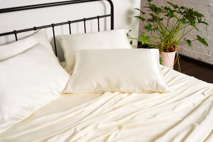
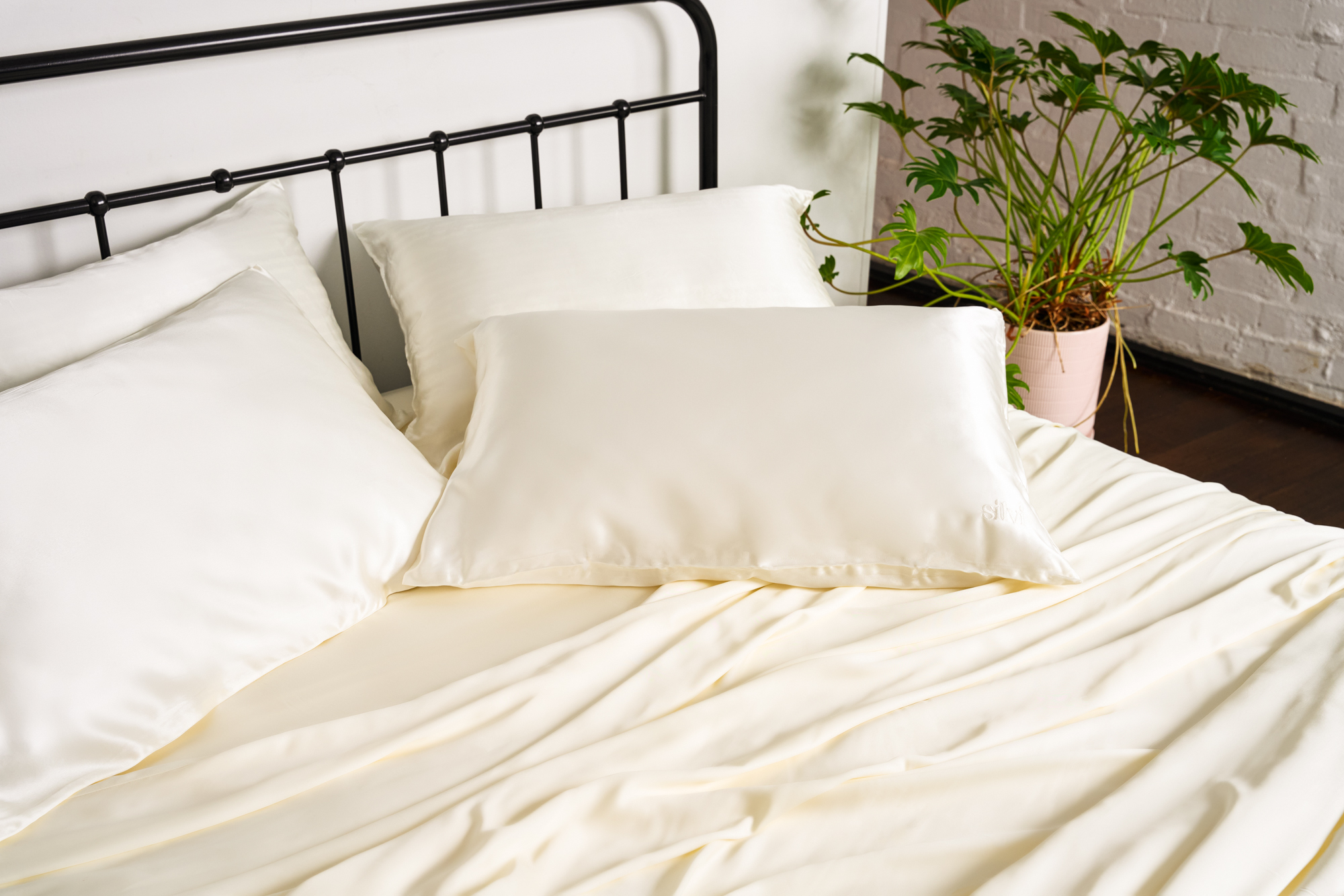
The Silvi silk pillowcase in white

ROSS CAMPBELL

The Silvi silk pillowcase in white

The Anti-Acne Silk Pillowcase comes in two sizes (standard and king size), as well as two colours (white and charcoal grey), to match your bedroom style. It also features a zip fastening so there’s no fear of your pillow slipping out.

Silvi’s pillowcase has been ethically certified cruelty free as it’s not tested on animals. And while the innovative design is machine washable – on a delicate setting with a neutral pH detergent – the silver treatment keeps it fresher for longer, so the pillowcase only needs washing every 10 to 14 days. Fewer washes means this purchase is more eco friendly, too. Every little helps.

Each pillowcase retails for £49, but fans say it’s an investment work making, considering the benefits to your hair, skin, sleep, and washing come laundry day. Buy for yourself, a friend, relative or loved one, and know that if you aren’t quite satisfied with your purchase, Silvi offers shoppers a 100-night guarantee, which means unsatisfied customers are eligible for a full refund.

But, with 100,000 pillowcases sold to happy customers in the first year alone, we don’t expect you’ll be using it any time soon. Sweet dreams!

Share Button

Help! My Toddler’s Taken A Serious Dislike To One Of Our Relatives

Having kids is all fun and games until your child decides they hate your relative – especially when you’re going to be spending Christmas with them.

One mum recently took to networking app Peanut to say her toddler had taken quite the dislike to her sister-in-law’s husband.

“She is a totally chill baby but when he comes over she will cry loads and cling to me,” she wrote. “I haven’t seen her like this with anyone else.”

What’s more, the mum said her sister-in-law’s husband is “the loveliest person”. What a pickle.

“I feel so bad that she doesn’t like him,” she wrote. “Any tips or advice? We’re having them over on Christmas Day and I’m worried. I also feel bad as she’s obsessed with my side of the family.”

There might be several reasons why little ones take a dislike to family members or friends, suggests Dr Kalanit Ben-Ari, a family therapist who founded the parenting platform Get The Village.

In some instances, a baby might sense a parent’s anxiety or stress around a particular person and react to it, she says. It might also be that once that person holds the baby, the child loses touch with the parents and becomes stressed, leading to fear of separation anxiety that is associated with that person.

“The problem is that when a parent anticipates the baby’s stressful reaction, the baby becomes more stressed, so it is a cycle or reaction that starts to be associated with that person,” says Dr Ben-Ari.

Everything a young child does – whether throwing themselves on the floor, or nuzzling their parents – is their way of demonstrating how they feel, because they often can’t verbalise it.

“In this instance, it sounds like the child is using lots of non-verbal messaging and non-verbal clues,” says Fiona Yassin, family psychotherapist and founder and clinical director of The Wave Clinic.

“Children often cry or cling to a parent in this way because they feel unsafe. It could be the person reminds them of someone else who looks quite scary,” she says.

The therapist offers the example that if the person has a beard, it could be that the child associates them with a bearded villain they’ve seen on TV and will remember the negative emotions they felt when they saw said villain – so “seeing this person may trigger a similar response”.

Sometimes children might have a sensory reaction to a family member that isn’t necessarily positive. They might take a dislike to a different voice, accent, smell or even appearance: for example, if they wear glasses or keep their shoes on in the house.

“Although these appear to be very small points, children look out for tiny differences, which is something we call the Little Professor,” says Yassin. “The Little Professor in the child has the job of trying to work out what’s going to happen next.”

What can parents do about it?

You’re probably dreading your child seeing the family member they’ve taken a dislike to, but you’re going to have to try really hard to think positively and anticipate a good connection. Otherwise your little one might pick up on your anxiety – fuelling the negative reaction further.

Ahead of seeing them, it might be helpful to create and share a positive persona of the person the child dislikes.

“If you have family photographs with the person in, show them to your child and talk about what that person does, who they are, who their family is and what activities they like to do,” says Yassin.

This helps the child to understand more about who they are and squash the idea of them as a villain-type character.

It can also be helpful to have your little one’s security blanket or toy on hand, suggests Dr Ben-Ari, to offer comfort and familiarity.

If the child’s reaction to the person is very strong, allow someone else to hold them while you greet and welcome the person, adds the therapist. “Seeing you at ease, relaxed and happy, interacting with that person will send the signal to the baby that it is safe.”

“Seeing you at ease, relaxed and happy, interacting with that person will send the signal to the baby that it is safe.”

– Dr Kalanit Ben-Ari

One thing you definitely shouldn’t do is force your child onto the person – whether that be letting them hug, kiss or hold them, which can ultimately just make things worse.

Instead, encourage the adult to give space to the baby to reach out to them, suggests Dr Ben-Ari. “The adult can start making fun noises, point to or hold a toy, offer the baby’s favourite toy, speak calmly to the baby, and once the baby shows signs of interest, they can slowly get closer.”

It’s also important not to leave your child alone with them – regardless of whether they’ve just arrived or have been there a while.

“When the person the child dislikes enters the room, it’s better they are not left on the floor or on their own,” says Yassin. “Hold the child or sit on the floor with them so you are physically at the same level.”

She continues: “Parents mustn’t force cuddles or plead the child to be nice to the person they have taken a dislike to. Railroading the situation and trying to force a relationship will create bigger barriers. Do not leave the child alone with the person for any length of time and be gentle with introductions.”

If the family – including the person the child dislikes – is coming for Christmas, keep things as normal as possible, says the therapist. And remember: it probably won’t last forever. “This type of situation tends to be a very big deal for a short period of time,” she adds, “but does blow over.”

Share Button

Ethereal color variant of mysterious plant is actually a new species

Green leaves and photosynthesis were once considered essential characteristics of plants. However, some plants have stopped performing photosynthesis and take the nutrients they need from other organisms instead. One such mycoheterotrophic plant is ghostly-looking Monotropastrum humile that is widely found across East and Southeast Asia. It often grows in woodlands where there is little sunlight, obtaining the nutrients it needs by feeding off the hyphae of fungi. Despite its wide distribution, it was previously believed that only one species of this plant existed in the world. However, Professor SUETSUGU Kenji and colleagues have discovered that a variant found in Japan is actually a new species, shaking up our understanding of this unusual-looking genus of plants.

It has rosy pink petals and stems resembling milk glass, giving it a beautiful, otherworldly appearance. As it was first found around Kirishima in Kagoshima Prefecture, Japan, the new species has been named Monotropastrum kirishimense.

Originally, this new species was tentatively treated as a color variant of M. humile, known as M. humile f. roseum. Thus began an extensive and multifaceted 20-year study to determine how exactly these plants differed. Specimens were collected from throughout Japan and Taiwan, as well as Vietnam.

Results of various analyses revealed morphological differences, including the following; M. kirishimense flowers and ovaries are more rounded than those of M. humile, and its rootball is more obscured by the surrounding soil (in contrast to M. humile‘s protruding root tips). M. kirishimense individuals are shorter above ground (under 5cm) and longer below ground (over 10cm). The flowering season is different too; M. humile flowers bloom approximately 40 days earlier than M. kirishimense. As the two plant species have the same primary pollinator (the bumblebee Bombus diversus), this difference in flowering times can reduce heterospecific pollen deposition, helping to ensure conspecific mating, and thereby preventing them from producing hybrids.

There are several other possible reasons why M. kirishimense and M. humile may have evolved into separate species. One possibility is that they have become specialized in feeding on different fungi, which has led to reproductive isolation, or the inability to produce offspring together. This process is known as resource partitioning and is one of the major ways that species can evolve from a common ancestor. Genetic analysis of mycobionts revealed that M. kirishimense has a consistent, specialized association with a particular lineage of fungi, whereas M. humile is associated with different lineages. Therefore, this study suggests that M. kirishimense may have evolved into a new species by relying on a specific type of fungus. In fact, the phylogenetic tree (a ‘family tree’ of the evolutionary history of a group of organisms) of the plants themselves shows that the genetic characteristics of M. kirishimense and M. humile can be separated into two clades. Based on the researchers’ analysis of various characteristics, it has been revealed that M. kirishimense is distinct from M. humile in terms of its appearance, flowering patterns, evolutionary history, and ecological relationships. Therefore, the researchers concluded that it should be recognized as an independent species.

Overall, the research group not only revealed that M. kirishimense is a distinct species but also deepened understanding of plants in the Monotropastrum genus. Mycohetrotrophic plants are very vulnerable to extinction as they rely on specific ecosystems to survive and are usually found in old-growth forests. The newly recognized species, M. kirishimense, is rare and presumably endangered. Now that it has been identified as a new species, conservation efforts can be made to protect it. This study emphasizes the importance of combining various analysis methods, called integrative taxonomy, to fully understand and protect biodiversity. This work was conducted by a multi-institutional research group, including Professor Suetsugu (Graduate School of Science, Kobe University), Professor SUYAMA Yoshihisa (Graduate School of Agricultural Science, Tohoku University), and Dr. Tian-Chuan Hsu (Taiwan Forestry Research Institute). The paper was published online in Journal of Plant Research on November 30, 2022.

Story Source:

Materials provided by Kobe University. Note: Content may be edited for style and length.

Share Button

Wafer-thin device has potential to transform the field of islet cell transplantation

A quarter-sized device created at Houston Methodist could drastically alter the course of treatment for Type 1 diabetes, a chronic condition that impacts millions of Americans and does not have a cure.

In a study published in the Dec. 26 issue of Nature Communications, a research team led by Houston Methodist delivered islet cells and immunotherapy directly into a 3D printed device akin to a bioengineered pancreas, called the NICHE. The treatment restored healthy glucose levels and eliminated Type 1 diabetes symptoms in animal models for more than 150 days while avoiding severe adverse effects of anti-rejection therapy by administering immunosuppressive drugs only where the transplanted islet cells were located.

Type 1 diabetes is caused by an autoimmune reaction that destroys the cells in the pancreas that make insulin. It can also cause kidney failure. Daily insulin injections are the most conventional treatment but attaining tight control of glucose levels remains challenging and cumbersome for patients. Further, in more severe cases, patients may need pancreas and kidney transplants, or they may qualify for an islet cell transplant, where the islet cells of a deceased pancreas donor are harvested, processed and then transplanted into the Type 1 diabetes patient’s liver.

These transplants can help improve a patient’s symptoms; however, as with all organ transplants, one of the biggest challenges is the need for immunosuppressive drugs for the rest of their lives to avoid transplant rejection. Lifelong immunosuppression can lead to patients being vulnerable to infectious diseases and increases the risk of certain types of cancer.

The NICHE, created in the Department of Nanomedicine at Houston Methodist Research Institute, is a flat device placed under the skin comprised of a cell reservoir for the islets and a surrounding drug reservoir for localized immunosuppression therapy. It is the first platform to combine direct vascularization and local immunosuppression into a single, implantable device for allogeneic islet transplantation and long-term Type 1 diabetes management. Direct vascularization is fundamental for supplying nutrients and oxygen for maintaining the viability of transplanted islet cells.

“A key result of our research is that local immunosuppression for cell transplantation is effective,” said Alessandro Grattoni, Ph.D., corresponding author and chair of the Department of Nanomedicine at Houston Methodist Research Institute. “This device could change the paradigm of how patients are managed and can have massive impact on treatment efficacy and improvement of patients’ quality of life.”

The NICHE incorporates ports for the refilling of drugs as needed. The researchers refilled the drug reservoirs every 28 days, which is comparable to other long-acting drugs clinically available for migraine prevention or HIV treatment.

Grattoni’s team is working on scaling up the NICHE technology for clinical deployment, for which drug refilling may only be needed once every six months. The ability to refill the NICHE technology allows for long-term use in patients. Further, changes in drug formulations or concentration could extend refill intervals to once each year, aligning with routine physician visits.

Grattoni and his collaborators will expand this research over the next few years, with the end goal of testing the NICHE’s safety in humans in about three years. Grattoni’s nanomedicine lab at Houston Methodist focuses on implantable nanofluidics-based platforms for controlled and long-term drug delivery and cell transplantation to treat chronic diseases.

Grattoni’s collaborators on this study include Jesus Paez-Mayorga, Jocelyn Nikita Campa-Carranza, Simone Capuani, Nathanael Hernandez, Hsuan-Chen Liu, Corrine Ying Xuan Chua, Fernanda Paola Pons-Faudoa, Gulsah Malgir, Bella Alvarez, Jean A. Niles, Lissenya B. Argueta, Xian C. Li, Joan E. Nichols and A. Osama Gaber (Houston Methodist); Kathryn A. Shelton, Sarah Kezar and Pramod N. Nehete (MD Anderson Cancer Center); and Dora M. Berman, Melissa A. Willman, Camillo Ricordi and Norma S. Kenyon (University of Miami).

This research received funding support from Juvenile Diabetes Research Foundation (1-INO-2018-595-A-N), Vivian L. Smith Foundation, Houston Methodist Research Institute, Diabetes Research Institute and the National Institutes of Health (NIDDK R01DK132104).

Story Source:

Materials provided by Houston Methodist. Note: Content may be edited for style and length.

Share Button