Small glowing protein allows researchers to peer deeper into living tissues

Biomedical and genetic engineers at Duke University and the Albert Einstein College of Medicine have designed a small fluorescent protein that emits and absorbs light that penetrates deep into biological tissue. Tailored to wavelengths in the near-infrared (NIR) spectrum, this protein can help researchers capture deeper, cleaner, more precise biomedical images.

This work appeared Dec. 1 in the journal Nature Methods.

Imaging deep tissues with light is challenging. Visible light is often quickly absorbed and scattered by structures and molecules in the body, preventing researchers from seeing deeper than a millimeter within a tissue. If they do manage to probe further, substances like collagen or melanin often muddy the image, creating the equivalent of background noise through their natural fluorescence.

“Biological molecules naturally absorb and emit light in the visible spectrum, which is about 350 to 700 nanometers,” said Junjie Yao, assistant professor of biomedical engineering at Duke. “So when using it to image deep tissue, it’s like trying to observe the stars in daylight. The signals get flooded out.”

To wade out from these muddied waters, Yao and his collaborator Vladislav Verkhusha, professor of genetics at Albert Einstein College of Medicine, New York developed a protein that absorbs and emits longer wavelengths of light in the near-infrared (NIR) spectrum.

“Tissue is the most transparent in the 700-1300 nanometer window of NIR light,” said Yao. “At those wavelengths, light can penetrate deeper into a tissue, and because there is less natural background fluorescence to filter out, we can take longer exposures and capture clearer images.”

Verkhusha and his lab used a process called directed molecular evolution to engineer their proteins, using photoreceptors normally found in bacteria as the basis for the structure. These photoreceptors are useful for imaging research because they can switch between a silent and active state when hit with a specific wavelength of light. They can bind with biliverdin, a biomolecule that appears in high quantities in mammalian and human tissues.

“We studied the structure of the biliverdin to determine how the photoreceptor would best attach to the biomolecule,” explained Verkhusha. “After we understood the binding process, we carefully introduced substitutions of key parts of the molecule connecting to the biliverdin to increase the electron binding, which is necessary to obtain the red-shifted fluorescence. Last, we performed random mutagenesis followed by high-throughput screenings so the proteins would evolve and increase their brightness.”

The brightest found protein, termed miRFP718nano, is easily produced in cells and tissues and emits light just outside of the visible range. But while the NIR activation itself is helpful, what happens after the initial burst of activity holds even more promise for biomedical imaging.

“We’ve seen that the NIR range can be broken up into two main zones,” explained Yao. “When NIR light first hit these proteins, they emit light in the first zone, which is about 700-900 nanometers. But as they decay, the wavelength gradually gets longer, like the tail behind a comet. That’s when they begin to emit light in the second NIR zone, which is from 900-1300 nanometers.”

In this second zone, all benefits from using the shorter wavelength, zone one NIR light are enhanced: light can penetrate tissue two times deeper, background fluorescence is significantly dimmed, and image resolution can be two to three times clearer, allowing for detailed images of complex structures.

As a proof of concept, Yao and his team at Duke used an imaging technique called short-wavelength infrared (SWIR) to test the efficacy of the new protein. This process sends NIR zone-one light deep into the tissue to activate the fluorescent proteins. As the proteins decay, they emit NIR zone-two light that provides information about the structure and composition of the targeted tissues and cells that can be translated into high-resolution images.

After introducing engineered miRFP718nano proteins into their animal models, the team used them to capture images of microbes in the mouse digestive tract, visualize cells in a mouse mammary gland and even track changes to inflammation in a mouse liver. All the images captured were clearer and more detailed than images made using a standard NIR zone-one imaging protein.

Yao and Verkhusha are optimistic that their continued partnership will be a boon for their work in both biomedical imaging and protein engineering. As Verkhusha continues to refine and improve the fluorescent proteins and biosensors, Yao is excited to use the new tool to visualize the brain more closely and to potentially track the movement of cancer cells.

“This is an exciting new front of our decade-long collaboration because we can use the imaging tools to guide the protein engineering decisions, and we can use the advanced protein engineering to improve the imaging capabilities,” Yao said.

This work was supported by grants from the US National Institutes of Health (GM122567, EB028143, NS111039, NS115581), the Chan Zuckerberg Initiative (226178), the Cancer Foundation Finland and Magnus Ehrnrooth Foundation, and the NIH Intramural Research Program for the Vaccine Research Center of the National Institute of Allergy and Infectious Diseases.

Share Button

Strep A death of eighth child confirmed

A child in Hampshire has died of strep A, officials confirm, bringing the total deaths to eight.

Share Button

Obstetrician Explains What Actually Happens Behind The Curtain During A C-Section

We all know vaguely what happens when you have a caesarean section – you’re given anaesthetic, a cut is made in your abdomen and then, minutes later, your baby is pulled out and passed to you. It’s a magical medical moment.

But it turns out there’s actually a hell of a lot going on from the obstetrician’s perspective. More than you could possibly know.

A fascinating Tiktok video explaining the anatomy of a C-section has been viewed more than 11 million times after showing just how complicated the surgery can be.

Using various sheets of coloured fabric and paper, Tina (@mamma_nurse) explained how there are various layers that surgeons have to cut through, before moving muscle out of the way and then manoeuvring past organs – and that’s before you’ve even reached the area where the baby is.

Most information given on C-sections by healthcare providers is lacking in details. Probably for good reason.

But if you’re the type of person who likes to be super informed ahead of birth – or you’re just really curious as to what the surgery involves – we asked Meg Wilson, an obstetrics and gynaecology consultant at London Gynaecology and the Whittington Hospital, to walk us through the process.

(Just a head’s up, there are some quite graphic images below.)

What happens during a C-section

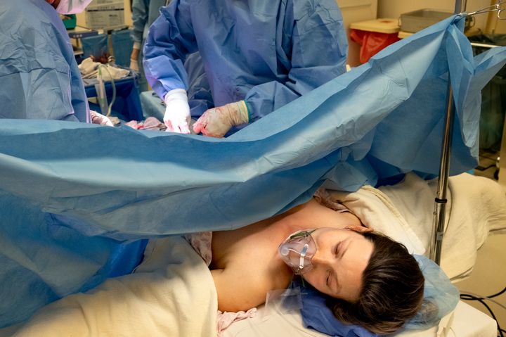
First up, you’ll be given some pain relief – either a regional or general anaesthetic – and a catheter is fitted. Your abdomen will be cleaned and a drape will be put up so you won’t be able to see the surgery unfold.

An obstetrician will make a 10 to 15cm cut along the skin at the bottom of your abdomen, just at the top of your pubic hairline.

The first layer they cut through is the skin, and that cut also goes through a layer of fat. “Then you come down on to the rectus sheath – a white fibrous layer – that is covering the muscles of the abdomen,” says Wilson.

They’ll make a cut in that as well and all these cuts will be done in the same direction: horizontally.

Byba Sepit via Getty Images

Next up, they come to the rectus muscles “which people know as their six pack muscles,” says the obstetrician.

These two muscles run as a strip down the abdomen and where they join together is something called the linea alba. It’s a weak area which surgeons can basically “poke through” to open it up, says the obstetrician.

“That takes you into your abdomen, by making that little hole, and then you’re into what we call the peritoneal cavity which is the proper wet inside of your tummy.”

Surgeons will make the hole a bit bigger by basically moving the two muscles apart.

“You’re pulling them out to the side to make a hole there,” says Wilson. “I think it’s really important that people know you’re not actually cutting muscles, you’re just shifting them apart.”

When in the peritoneal cavity, surgeons will get a nice view of the surface of your bladder and then the main event: the womb itself.

Now comes the really interesting bit. They have to actually move organs to get to where they need to be – so yes, they shift your bladder out of the way.

“You lift up a little bit of something called the peritoneum,” she explains, which is “like a sheet that runs over the womb and the bladder.” Surgeons will make a small hole in that “sheet” and this allows them to see where the bladder is attached to the womb.

“It allows you to push the bladder out of the way and push it downwards a little bit,” Wilson says, noting it only gets moved about 1-2cm.

In the operating room, they have a specially designed surgical instrument that goes in to protect the bladder and hold it down and out of the way.

The surgeon cuts into the womb (again, a horizontal cut in the same direction as the skin cut). The womb is a muscle so they expect to have some bleeding at this point as muscles have a strong blood supply.

In cases where a parent has placenta previa – where the placenta completely or partially covers the opening of the uterus – it might be a bit more complicated, she adds, and they might have to cut higher up in the womb or even through the placenta, which could result in more blood loss.

“Then you’re going to hit the membranes around the baby – the amniotic sac – and you see that bulging out when you reach it,” Wilson continues. “You keep going and make a little hole in that with the scalpel and usually lots of amniotic fluid all comes spilling out in a big gush.”

At this point it’s all about getting the baby out. In a straightforward pregnancy the baby will be in a head down position, so they’ll get a nice view of that.

The retrieval process involves a bit of teamwork. “You put your hand in and slip your hand around the baby’s head – like a cupping action – and your assistant will put lots of pressure on the top of the woman’s tummy, pushing right at the top of womb where the baby’s feet are, and you’ve got your hands acting as a little slide for the baby to come out,” she says.

The head pops out, then comes the neck, and then there’ll be a bit more gentle pulling to deliver the shoulders one at a time, and then the body “slips out relatively easily after”.

Doctors work to remove a baby from a woman's uterus during a c section.

Michael Hanson via Getty Images

Doctors work to remove a baby from a woman’s uterus during a c section.

At this stage the curtain is often lowered and the parent(s) can see their baby and hold them. They tend to let the umbilical cord pulsate for at least a minute to give the baby beneficial nutrients and then they’ll clamp the cord, cut it and hand the baby to the midwife who will wrap them up and dry them off.

“Then it’s a case of removing the placenta, which is still stuck on to the lining of the inside of the womb,” says Wilson. “We pull a little bit on the cord and the placenta will be detaching.”

After the placenta has been removed, surgeons will then clean out the inside of the womb which Wilson says is “a very simple action” of rubbing a swab around the insides of the cavity “to make sure there’s no little bits of membrane or last little threads of placenta that are still there”.

The mum will still be bleeding at this point so time is of the essence to get the womb stitched up again. Once that’s done, surgeons will take a moment to do a check and assess that there’s no additional bleeding anywhere.

bymuratdeniz via Getty Images

At this stage, she says, they clean away any spilt fluid and blood that may have collected in the sides of the pelvis using a swab. They also check the womb is contracted and that the ovaries and fallopian tubes look normal.

“It’s just an opportunity to do a health check because you’ve got the tummy open and it’s a relatively straightforward thing to do,” she adds.

The instrument protecting the bladder is then taken out and “then you allow everything to fall back into place,” adds Wilson. “The bladder will go back to where it was originally placed and those two rectus muscles that you held open to get into the cavity of the tummy will fall back into place as well.

“You don’t routinely close the rectus muscles … they will come back together naturally and reform their meeting point.”

The next layer that’s surgically closed is the rectus sheath, which is the layer just beneath the fat. “We sometimes put a stitch in the fat layer but for most women we then just close the skin with another running stitch,” she adds.

“By routine we remove all the drapes and clean any collected blood that’s in the vagina,” she adds, “and make sure there’s no clots of blood or anything.”

At this point they might put in a painkiller suppository and a dressing is placed over the c-section wound. The whole process is complete in less than an hour.

Share Button

How To Pick A Workout Playlist That’ll Keep You Going For Longer

You’re reading Move, the nudge we need to get active, however makes us happiest and healthiest.

Music is a lot of things: it’s restorative, motivational, moving and educational. There are endless ways we use music to get through our days, whether listening to a sad song on repeat or hitting play on an upbeat tune.

According to Ronna Kaplan, a clinical supervisor and adjunct music therapy faculty at Cleveland State University, “music is positive in many ways for mental health, it can be used across the lifespan” for many different situations.

One of those ways is during exercise. It can be a crucial element in enhancing your workout. Here’s how.

Your body’s movement naturally matches a song’s rhythm, which can help you stick to a specific pace.

There’s a reason your foot starts tapping or your shoulders start moving as soon as a song comes on. According to Joy Allen, the chair of music therapy and director of the music and health institute at Berklee College of Music in Boston, this is because of rhythmic entrainment, which is an “unconscious reaction — that’s what we call the entrainment.”

“Our body’s going to [move] in time with that sound or that rhythm,” she says.

So, when it comes to exercise, your body automatically falls in line with the tempo of the music “because of the way that our brains are connected with rhythm,” Allen says.

When picking music for a workout, like when going for a walk or run, for example, you’ll want to choose a tempo that is close to your natural stride. “Go [with] what seems comfortable for you and play around with different songs,” she says.

You can use music to increase your pace, too.

If you’re looking for an added challenge, pick a song with a pace that is a little quicker than your average running or walking stride, this should help you move faster throughout your workout.

You can start with a song with a slower tempo and gradually increase your speed by picking songs with faster beats, which is ideal if you’re looking to improve your walking or running pace, according to Kaplan.

“It primes the person to an outside cue,” she says. It “helps your muscles activate in their walking pattern.”

Music can help distract you from boredom during a workout.

Tempura via Getty Images

Music can help distract you from boredom during a workout.

It’s motivating.

How often has someone walked into the gym, realised they forgot their headphones, and then had a not-so-great workout — or even left the gym altogether? Allen pointed out this is a common occurrence: There is a major reason why music is integral to so many people’s workouts.

The music you listen to during a workout helps with motivation, and there are several things behind that motivation.

First, you probably want to hear your favorite song on your exercise playlist, which may keep you going for longer. Second, if you put on music that’s unexpected (like if you put on reggaeton instead of your regular pop soundtrack), you will be interested in hearing what comes next in the song, which may also keep you moving longer than usual.

“If you’re always listening to the same stuff, sometimes that’s great [but] sometimes we have fatigue from it — we know what to expect and what’s coming, so it can be a little less motivating,” Allen says.

And music is distracting.

No one wants to focus on their tough workout as they’re in it. If anything, they want to not think about it. As you sing along to lyrics or are reminded of music-induced memories, songs let your mind wander throughout an exercise regimen, so you don’t have to stand (or sit) there and think about how hard your workout is.

Music keeps you from getting bored during a workout, too, which can happen when you’re doing something kind of mundane like walking on a treadmill, Allen noted. Music activates the brain by giving your mind something else to think about.

“It captures your attention… ‘oh, here’s something I’m listening to,’ so I’m not attending to what could be an uncomfortable experience with the exercise, it gives me something else to focus on,” Allen says.

You’ll reap even more benefits when you pick your music.

According to Kaplan, when someone chooses the music they’re listening to, they’ll have better results, whether working out or doing something like meditation.

A recent study led by the Department of Kinesiology at Samford University in Alabama stated, “if the music played over the speakers is not preferred by the individual giving effort, performance may suffer. Thus, coaches and athletes should consider individual music preferences when attempting to optimise performance and training.”

This further speaks to the motivation you feel when working out to music you enjoy.

Additionally, Kaplan says you might notice you’re in a better mood when working out to music you select, which may make you feel like you enjoyed your workout more. And that’s a win-win.

This may mean you’ll be more likely to work out again that week, which is a great way to hit your fitness goals.

Move celebrates exercise in all its forms, with accessible features encouraging you to add movement into your day – because it’s not just good for the body, but the mind, too. We get it: workouts can be a bit of a slog, but there are ways you can move more without dreading it. Whether you love hikes, bike rides, YouTube workouts or hula hoop routines, exercise should be something to enjoy.

HuffPost UK / Rebecca Zisser

Share Button

Strep A: Penarth family traumatised by daughter’s death

The father of seven-year-old Hanna Roap, describes how his “bubbly”daughter died within hours.

Share Button

Accelerating pathogen identification in infants and children with bloodstream infections

A collaborative team led by researchers from Great Ormond Street Institute of Child Health (GOSH), London and including researchers from the Wyss Institute for Biologically Inspired Engineering at Harvard University and BOA Biomedical in Cambridge has re-engineered the process of microbial pathogen identification in blood samples from pediatric sepsis patients using the Wyss Institute’s FcMBL broad-spectrum pathogen capture technology. The advance enables accurate pathogen detection with a combination of unprecedented sensitivity and speed, and could significantly improve clinical outcomes for pediatric and older patients with bloodstream infections (BSIs) and sepsis. The findings were published in PLoS ONE.

BSIs with various microbial pathogens can rapidly escalate to life-threatening sepsis when the body is overwhelmed by the multiplying invaders and shuts down its organs’ functions. In 2017, there were 48.9 million cases and 11 million sepsis-related deaths worldwide. Importantly, almost half of all global sepsis cases occurred among children, with an estimated 20 million cases and 2.9 million global deaths in those under five years of age.

To prevent BSIs from progressing to full-blown sepsis, the infection-causing bacterial or fungal species must be identified as fast as possible. Only then can optimal pathogen-tailored antibacterial or antifungal treatments be applied in time. The conventional method used in clinical laboratories to identify the causative pathogenic species is long and laborious, requiring two time-consuming culture steps that take at least 1 to 3 days to complete.

“For all patients with sepsis, their chances of surviving dramatically shrink the longer it takes to identify the infection-causing pathogen(s) and thus, receive the most promising antimicrobial treatment,” said Nigel Klein, M.D., Ph.D., pls, a Professor of Infectious Disease and Immunology at GOSH, and a senior author on the study. “At Great Ormond Street Hospital we have been working to demonstrate both the importance rapid diagnosis and the fact that with innovative approaches we can identify the causative organism in between 40 minutes and six hours. Compared to adult patients, sepsis in infants and small children progresses much faster, and therefore there is a real need for diagnostic methods that support early detection. Accurate diagnosis is even more significant due to the availability of only small blood volumes from paediatric patients which can make re-sampling challenging.”

In 2020, senior authors Klein and Elaine Cloutman-Green, Ph.D., a Consultant Clinical Scientist and Infection Control Doctor at GOSH, began collaborating with Lead Staff Scientist Michael Super, Ph.D. and Founding Director Donald Ingber, M.D., Ph.D. at Harvard’s Wyss Institute to solve this problem. “Based on our earlier success with FcMBL in isolating pathogens from joints as well as bovine and human blood with extraordinary efficiencies, we hypothesized that building an FcMBL-mediated pathogen capture into a modified clinical blood culture protocol could shorten the time and reduce the size of the required patient samples to yield the same results that time-consuming blood culture protocols provide,” said Super.

In the pathogen identification process currently performed in clinical settings, first, blood samples are added to bottles containing liquid media in which infectious microbes, if present, are amplified to a certain density. Then, the amplified microbes are grown on solid media as isolated colonies whose constituent cells eventually can be identified with a highly sensitive, yet fast and relatively inexpensive analytical method know as MALDI-TOF mass spectrometry (MS). “Indeed, isolating the infectious microbes directly from grown liquid blood cultures using FcMBL makes them available for MALDI-TOF MS analysis much earlier,” added Super.

FcMBL is the key component of a broad-spectrum pathogen capture technology. It consists of a genetically engineered human immune protein called mannose-binding lectin (MBL) that is fused to the Fc fragment of an antibody molecule to produce the resulting FcMBL protein. In this configuration, the MBL portion of FcMBL can capture more than 100 [CHECK WITH MIKE] different microbial species with high efficiency, including virtually all of the bacterial and fungal pathogens causing sepsis. FcMBL’s Fc portion can be used to couple it to magnetic beads, allowing the captured pathogens to be quickly pulled out of patient samples and liquid blood cultures.

In the earlier stages of the project, the Wyss team provided purified bead-coupled FcMBL to the GOSH team, which had access to blood samples from pediatric patients at the hospital. At later stages, the sepsis and infectious disease company BOA Biomedical, co-founded by Super and Ingber to commercialize the Wyss Institute’s FcMBL technology, provided the FcMBL reagent and critical expertise to the project. BOA Biomedical meanwhile developed the manufacturing capabilities for FcMBL that the Food and Drug Administration (FDA) in the US and other federal health agencies require for producing therapeutic and diagnostic products.

“Sepsis is the leading killer in hospitals, and rapidly initiating the right antibiotic saves lives. Using work originally developed at the Wyss Institute, BOA Biomedical’s revolutionary FcMBL technology helps to quickly and accurately identify the pathogen causing sepsis, ushering in a new era of targeted antimicrobial therapy to help individual patients and curb society’s deadly antimicrobial resistance problem,” stated Mike McCurdy, M.D., Chief Medical Officer of BOA Biomedical.

In addition to using the gold standard two-step blood culture in combination with MALDI-TOF MS pathogen identification, the team also included the Bruker Corporation’s MBT Sepsityper® kit as a comparison. Brought to market in 2021, the MBT Sepsityper® essentially eliminates the time-consuming second microbial culture step by lysing microbial cells from the liquid culture and spinning the fragments down in a centrifuge before analyzing them by MALDI-TOF mass spectrometry analysis. Although it accelerates the overall diagnostic process, the MBT Sepsityper® methodproduces lower microbial detection rates than those obtained with the conventional culture method, which means that it may still fail to identify the infection-causing pathogen in a significant fraction of blood samples.

“Our FcMBL approach has opened up the opportunity to identify pathogenic organisms to guide treatment 24 to 48 hours earlier than would be possible using standard culture techniques. It has also enabled us to use this identification to make any ongoing culture for antibiotic sensitivities more tailored to the needs of the patient. This method isn’t tied into a specific platform or manufacturer, and thus we see clear potential for it to become a new standard processing step for clinical pathogen detection,” said Cloutman-Green.

“The FcMBL method identified 94.1% of microbial species found in clinical blood culture analysis with samples from 68 pediatric patients,” said first author Kerry Kite, who performed her graduate work with Klein and Cloutman-Green. “We were able to identify more infectious species in positive liquid blood cultures using the FcMBL method than with the MBT Sepsityper® method (25 of 25 vs 17 of 25), and this trend was even further pronounced in the case of the common fungal pathogen Candida (24 of 24 vs 9 of 24).” Candida species account for about 5% of all cases of severe sepsis and are the fourth most common pathogen isolated from patients’ bloodstreams in the United States. Not only do infections with Candida and other fungi require specific antifungal treatments, distinguishing among the various types of pathogenic fungi helps direct the appropriate antimicrobial therapy. Specifically in neonatal intensive care units, Candidainfections are a major cause of morbidity and mortality, killing as many as 40% of infants and often causing neurodevelopmental impairments in those that survive.

“By continuously adapting the powerful FcMBL pathogen capture technology to unmet and pressing diagnostic needs, such as the rapid diagnosis of sepsis in pediatric patients, we hope to profoundly alter the frequently dismal prospects of patients of all ages,” said Ingber. “Our ultimate goal is to be able to accurately and even more rapidly identify pathogens directly in small samples of blood without the need for any additional microbial cultures.” Ingber is also the Judah Folkman Professor of Vascular Biology at Harvard Medical School and Boston Children’s Hospital, and the Hansjörg Wyss Professor of Bioinspired Engineering at the Harvard John A. Paulson School of Engineering and Applied Sciences.

The study was also authored by Sahil Loomba and Thomas Elliott at Imperial College London; Francis Yongblah, Lily Gates and Dagmar Alber at GOSH; George Downey and James Hill at BOA Biomedical; and Shanda Lightbown and Thomas Doyle at the Wyss Institute. The authors were supported in their work by the clinical microbiology staff at GOSH, as well as Erika Tranfield with MALDI-TOF MS expertise. At GOSH, critical financial help for the project from the Benecare Foundation, philanthropists Luca Albertini and Professor Pauline Barrieu, as well the Office of Vice-President (Advancement) at University College London was coordinated by Simona Santojanni. At the Wyss Institute, the study was funded by the Defense Advanced Research Projects Agency (DARPA) under Cooperative Agreement Number W911NF-16-C-0050, and the Wyss Institute’s technology translation engine. Additional support was provided by BOA Biomedical.

Share Button

Elf On The Shelf: 7 Simple(ish) Ideas To See You Through The Week

Admittedly some parents are more into it than others, going to great lengths to build sets involving their naughty elves. Some have even created Instagram accounts devoted to the mischievous elves running riot in their homes.

Meanwhile others are a little more laidback about the whole affair.

If you’re stuck for inspiration, we’ve scoured Instagram to find some relatively simple – but still creative – elf on the shelf ideas.

1. Frying eggs

2. Mummified

3. Grating a snowman’s nose

4. Doodling on bananas

5. Watching the World Cup

6. Making snow angels

7. Causing chaos in the loo

Share Button

‘Better Off Hibernating’: What It’s Really Like To Live With Seasonal Depression

While many of us may catch a case of the winter blues as the days get shorter, an estimated 10 million adults in the US and 2 million in the UK live with seasonal affective disorder.

This mental health condition, also known as seasonal depression, lasts around four to five months on average. “The appropriately named SAD, or seasonal affective disorder, is considered a type of depression characterised by its seasonal nature,” Jeff Temple, a professor at University of Texas Medical Branch and a licensed psychologist, tells HuffPost.

Unsurprisingly, people living farther north with less sunlight and earlier sunsets are more likely to experience SAD, likely due to the lack of sunlight and much shorter days. However, that doesn’t mean people in warmer, sunnier climates aren’t affected. While seasonal depression is much more common during the winter months, some people may experience seasonal depression during summer or during the colder season because of the fewer daylight hours. Women experience it more frequently than men.

A common misconception is that SAD is less serious than major depressive disorder because it doesn’t last all year round. However, people with this mental health condition explained to HuffPost that the symptoms of SAD are very real, and can even be debilitating.

Here’s what they want you to know:

People with seasonal depression may struggle to do daily activities

Like those with major depressive disorder, people with SAD may experience a lack of motivation and a loss of interest in day-to-day activities.

“I have had SAD for about 12 years, but I didn’t recognize the pattern or be diagnosed until about five years ago,” Claire, an optometrist, tells HuffPost. (Claire, along with some others in this story, asked to keep her last name private so she could freely talk about her mental health condition.) “I am typically an early riser and consider myself a productive person. However, when SAD hits, I struggle to do even the smallest daily chores like doing the dishes or making dinner.”

Similarly, Rebecca, a grad student, says that she has a “difficult time keeping up with [her] self-care and workout routine during the winter months,” and finds it nearly impossible to “function at full capacity.”

People with SAD may also feel more isolated during the darker months

Moreover, staying socially and emotionally connected to loved ones can require much more effort when coping with seasonal depression.

“The most prominent SAD symptoms I experience are loneliness and apathy. I tend to become quite numb in the winter months, and feel emotionally separated from the people around me,” says Vera, a freelance illustrator. “During regular depressive episodes, I may be able to ‘mask’ for most basic social interactions, whereas in winter seasons it’s not even an option. I’m too wiped out to even show up or pretend.”

Seasonal depression can interrupt normal sleep cycles and lead to extreme fatigue

SAD and sleep disturbances go hand in hand. Chloé Perrin, a bartender, has found that her symptoms of seasonal depression often manifest as constant exhaustion, leading to hypersomnia. Simply put, hypersomnia is characterised by recurring episodes of sleepiness during the day, difficulty waking up in the morning, and feeling tired despite oversleeping.

“My family used to joke that every winter I’d hibernate, whereas my sleep is otherwise normal-to-low the rest of the year and during other episodes,” she says.

Bella Sutter, a dancer with seasonal depression, explains that “getting out of bed feels impossible and my mornings normally start late.”

“I feel as if I would be better off hibernating through the winter because seasonal depression makes me feel like I’m half asleep anyway,” she adds.

SAD can cause mood changes, sleep disruptions and more.

Krzysztof Krysiak / EyeEm via Getty Images

SAD can cause mood changes, sleep disruptions and more.

People with seasonal depression may experience changes related to appetite and eating habits

Not only does seasonal depression impact sleep, but this mental health condition can have a negative impact on a person’s eating habits. Researchers have found a marked correlation between SAD and higher rates of disordered eating, including behaviours such as binging, purging and restricting food intake.

For Anna Samanamú, a high school paraprofessional and graphic designer, symptoms of SAD began when she was a teenager. “My appetite becomes affected [with SAD], and I would prefer to sleep rather than eat,” she says. “Unfortunately, that led me to have issues such as anaemia and Vitamin D deficiency.”

Seasonal depression may worsen symptoms of other mental health conditions

According to Temple, “seasonal affective disorder has a bi-directional relationship with other mood disorders like depression and bipolar disorder — meaning that people with one form are much more likely to develop or worsen the other.” Notably, SAD affects up to 20% of people with major depressive disorder, and 25% of people with bipolar disorder.

Rhiannon Bellia, who works in social services, has found that seasonal depression tends to exacerbate symptoms of other mental health conditions.

“My OCD gets a lot worse in the winter, it’s harder for me to focus and gauge time with my ADHD. With having autism, it’s also a bit harder for me to regulate sensory overwhelm,” Bellia says.

There are treatments for SAD

Temple explains that if symptoms such as losing interest in activities or feeling sad last “longer than a couple of weeks or [start] to interfere with your work, family, or relationships, then that’s a good sign that you may need some extra help.”

Fortunately, light therapy and cognitive behavioural therapy are proven to be effective treatments for people with SAD. The most beneficial at-home light therapy boxes provide light at 10,000 lux.

SAD is not simply feeling a little gloomy when the sun goes down; it is a serious mental health condition that can greatly impact every aspect of someone’s life.

“Please be kind to everyone who struggles with seasonal depression,” Samanamú says. “We are not lazy — sometimes there is just a disconnect from our minds and bodies. Many of us just need to take a little bit of time to become whole once again.”

Help and support:

  • Mind, open Monday to Friday, 9am-6pm on 0300 123 3393.
  • Samaritans offers a listening service which is open 24 hours a day, on 116 123 (UK and ROI – this number is FREE to call and will not appear on your phone bill).
  • CALM (the Campaign Against Living Miserably) offer a helpline open 5pm-midnight, 365 days a year, on 0800 58 58 58, and a webchat service.
  • The Mix is a free support service for people under 25. Call 0808 808 4994 or email help@themix.org.uk
  • Rethink Mental Illness offers practical help through its advice line which can be reached on 0808 801 0525 (Monday to Friday 10am-4pm). More info can be found on rethink.org.
Share Button

We Love Oodie’s Super Soft Wearable Blankets, Especially When They’re On Sale

We hope you love the products we recommend! All of them were independently selected by our editors. Just so you know, HuffPost UK may collect a share of sales or other compensation from the links on this page if you decide to shop from them. Oh, and FYI — prices are accurate and items in stock as of time of publication.

Picture this: it’s the Christmas break and you’ve finished work or studies for the holidays – or maybe just the weekend. It’s finally snowing (yep, we might get actually get a White Christmas this year) and you’re sprawled out at one end of the sofa enjoying a festive movie marathon with your BFF or partner curled up on the other, with no intention of moving for hours, unless it’s to dip your hand into a new chocolate selection box.

Perfection? Almost. There’s just one thing missing – an Oodie (or two).

If you’ve not heard of Oodie – aka “the world’s leading wearable blanket” – it’s the ultimate wardrobe must-have this winter. Ok, ok, so it might not be high fashion, but as our heating bills skyrocket, there’s a reason this brilliant hybrid of fleece, blanket, jumper and dressing gown is proving quite so popular.

Essentially, it’s a wearable hug, and perfect for anyone looking for a practical and comfy way to keep snug and warm this winter. And here’s the best part: Oodie is currently offering shoppers the chance to buy an Oodie for you and your favourite person – in the form of the discounted Oodie Twin Pack.

Oodie

An Oodie comes in just one, oversized size – based on a 6XL – which not only makes for the ultimate comfort and coverage, but also super simple shopping.

It boasts an insulating sherpa fleece lining, as well as an ultra soft exterior of Toastyek flannel fleece, and it’s this magic combo that will give you the warm and fuzzies. This design also features a giant front pocket to protect your hands from the chill, not to mention store your valuables and snacks, and an all important hood to keep your head and neck protected from any draughts. It’s also cruelty free and machine washable, making light work of laundry day.

The Oodie is available in over 40 colourways, including a variety of patterns from foodie to pet prints, tie dye, even your starsign – as well as plain block colours if that’s more your thing.

Not all patterns are included in the deal, fyi, so do double check before you buy. But whether you wear your Oodie to run errands, on a duvet day, working from home, or on winter walks, it’s the one. And now’s your chance to buy two!

Oodie twin pack
Oodie twin pack

The Oodie Twin Pack has been reduced from £178 to just £98, meaning you’ll be spending just £44 per Oodie in this deal. A single Oodie usually retails for £89 (or £69 on sale) so you’re practically getting two for the price of one here.

What we also love about this bundle is that you can select two different Oodie designs – allowing you to mix or match depending on your vibe. Personally, we’re going for pizzas and koalas in our bundle, but that’s how we roll.

This two-pack discount has already been applied on site, but, to claim it, shoppers must enter the discount code “CUSTOM-TWIN” at the checkout.

The deal coincides with Oodie’s Holiday Sale, with select other items on site reduced by £25, as well as great deals on family bundles and six-pack sets if you want to co-ordinate your entire friendship group or family, including your pets. The Kid’s Oodie is also on sale for just £52 in the run up to Christmas!

Fans of the brand can also shop its weighted blanket to warm the cockles this season, as well as the Oodie Robe, pyjamas, hair wraps, slippers and more. As always, these deals won’t last forever, so move quickly to bag a bargain.

Share Button

Father of girl, 4, fighting for life with Strep A infection is ‘praying for a miracle’

Dean Burns urges other parents with any concerns about their children’s health to get them checked.

Share Button